
Presentation
Two months history of dry cough, shortness of breath and weight loss (but no history of fever).
Patient Data
Age: 15 years
Gender: Female
From the case:
Ewing sarcoma

Download
Info
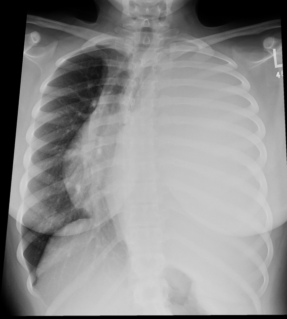
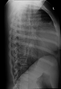
Demonstrates complete opacification of the left hemithorax, with marked positive mass effect and shift of the mediastinum towards the right.
Download
Info
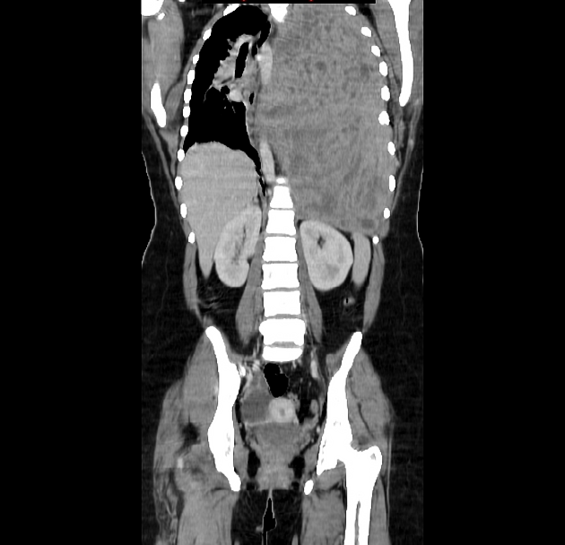
CT demonstrates a very large heterogeneous mass which appears to be confined to the chest, although the extra-pleural fat plane cannot be identified in some places.
Case Discussion
Tumor represents a Ewing sarcoma of the chest wall (formerly known as Askin tumor or pPNET) - see Ewing sarcoma family of tumors.


 Unable to process the form. Check for errors and try again.