
Hepatic leiomyosarcomas are rare primary malignant tumours derived from smooth muscle cells in the liver.
On this page:
Epidemiology
Hepatic leiomyosarcoma is rare 1. An equal sex distribution and a broad age range (5 months-66Y) has been reported. Some have suggested an associated with AIDS 2.
Pathology
The tumour may derive from smooth muscle cells of the bile ducts or blood vessels. No underlying aetiologic factors are known. Not associated with cirrhosis, but may be related to EBV. C-kit negative (differentiates it from GIST).
Microscopic features
Uniform spindle cells with blunt nuclear ends. Variable mitoses. A few large cells may be noted.
Immunophenotype
Vimentin and desmin positive.
Radiographic features
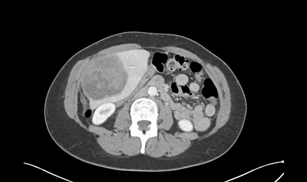
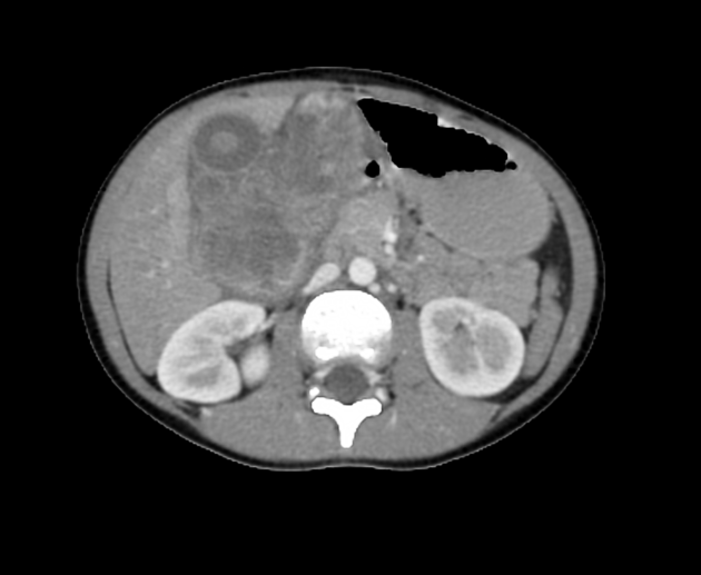
CT
"pseudocystic pattern" with heterogeneous enhancement 3
commonly large at diagnosis (6-35 cm)
may be predisposed to rupture/haemorrhage 4
pedunculated leiomyosarcoma has been reported and may arise from the ligamentum teres
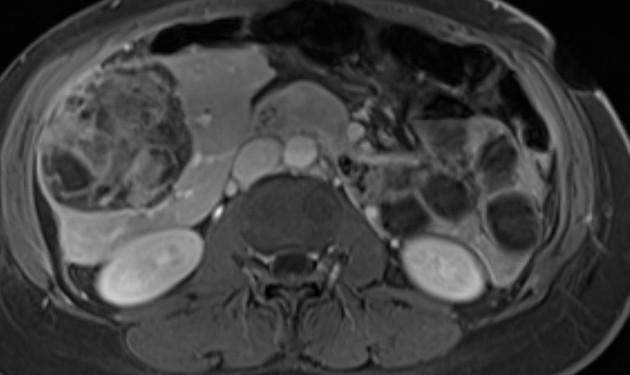
MRI
relatively well-marginated mass
heterogeneous T2 signal
intratumoural haemorrhage may result in patchy increase in T1 signal
slowly increasing enhancement on later postcontrast dynamic phases
foci of restricted diffusion
PET-CT
markedly hypermetabolic
Differential diagnosis
metastatic nonhepatic leiomyosarcoma (e.g. uterus, gastrointestinal, etc)
fibrosarcoma
malignant fibrous histiocytoma
solitary fibrous mesothelioma
Treatment and prognosis
Surgical resection is the standard treatment but with the rarity of the tumour, its effectiveness is uncertain. It had previously been reported that the majority of patients have recurrent tumour and some develop widespread metastatic disease, however, some studies suggest that this prognosis was based on some earlier misclassification of GIST tumours as hepatic leiomyosarcomas 5,7. The clinical course of a pedunculated leiomyosarcoma may be benign 6.


 Unable to process the form. Check for errors and try again.