
Presentation
Right upper quadrant pain for two months
Patient Data


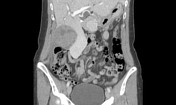
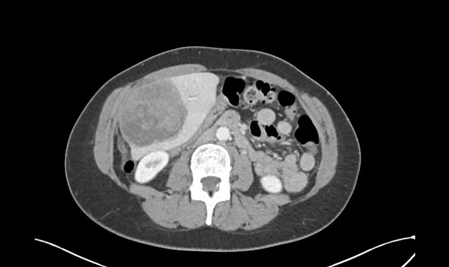
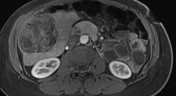
Heterogeneous attenuation 14 cm mass in the right hepatic lobe, with predominantly low attenuation. There is mild, patchy enhancement in the lesion.
Stranding at the anterior, inferior surface of the liver, extending into the omentum and peritoneal space is suggestive of rupture.
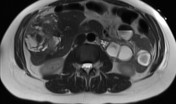
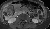


An MRI of the abdomen shows a Heterogeneous mass in the right hepatic lobe. It has a relatively well-defined capsule. On the T1W precontrast sequence, there are regions of hyperintensity within the mass, compatible with intra-tumoral hemorrhage.
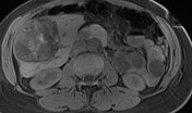
On the early arterial phase, the mass does enhance avidly, and T1 hyperintense regions in the mass are compatible with intratumoral blood products. There is mild abnormal enhancement on the anterior surface of the liver, compatible with tumor rupture.
On the portal venous phase in the dynamic postcontrast sequence, septa within the mass began to enhance, and there is the appearance of an enhancing capsule. Again seen is abnormal enhancement at the anterior surface of the mass, extending into the peritoneal space, compatible with tumor rupture.
Foci of restricted diffusion in the mass.

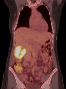
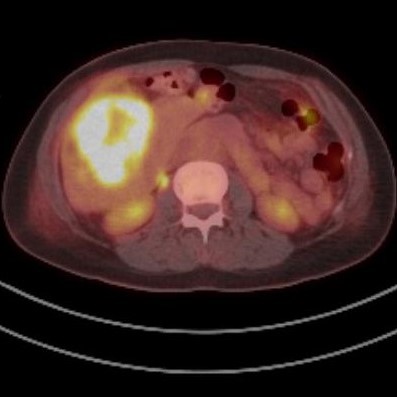
The hepatic lesion is markedly hypermetabolic, with an SUV of 10.2 (background liver is 1.4).
Case Discussion
Hepatic leiomyosarcoma is a rare primary tumor, with approximately 30-50 cases recorded. The tumor is thought to arise from smooth muscle cells in bile ducts or blood vessels, and has not been associated with cirrhosis.
The case above was resected (right hepatectomy) following the imaging findings. When the tumor was exposed, frank perforation and hemorrhage was noted, concordant with the images above (this finding has been described in other hepatic leiomyosarcomas). There was no carcinomatosis.
Pathologic analysis revealed a leiomyosarcoma penetrating through the liver capsule.
This case may be the first to demonstrate a hepatic leiomyosarcoma on PET-CT. Of note, the marked metabolic activity appears slightly discordant with the lack of avid enhancement in the tumor.


 Unable to process the form. Check for errors and try again.