Citation, DOI, disclosures and article data
Citation:
Bell D, Walizai T, Sharma R, et al. Intramural fat of the urinary bladder. Reference article, Radiopaedia.org (Accessed on 01 Mar 2025) https://doi.org/10.53347/rID-65168
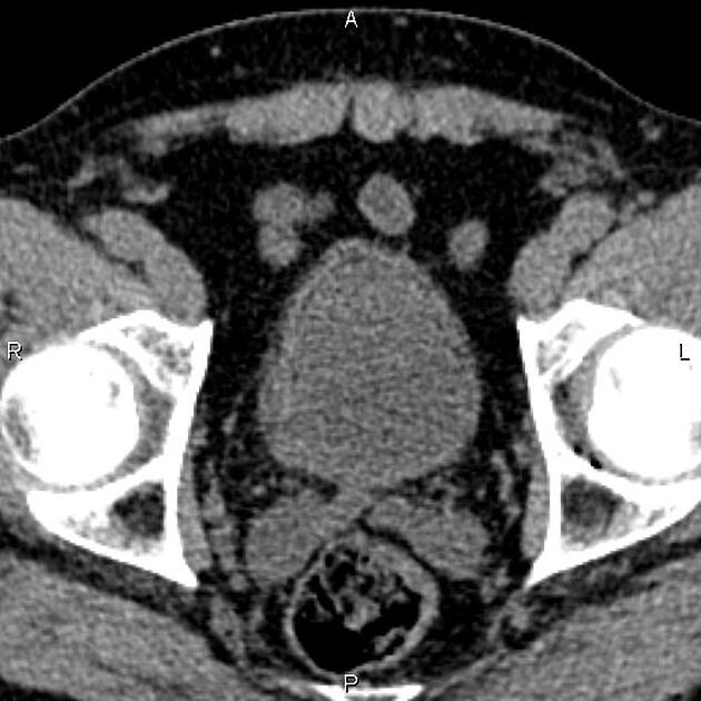
Intramural fat of the urinary bladder is an occasional benign finding on CT/MRI.
The incidence of this finding in histopathological studies is up to 4%. There is a male predominance 4.
It is typically an incidental, asymptomatic finding.
Adipocytes are found both within the lamina propria and the muscularis propria components of the wall of the urinary bladder 3.
On CT, a thin fat density stripe is visible along the bladder wall, primarily at the dome and anteriorly. It is more commonly seen when the bladder is only partially filled with urine 2.
It should not be confused with emphysematous cystitis, in which bubbles of gas are present within the wall. No lipid-fluid level is visible within the bladder proper (cf. chyluria).
-
1. Kriegshauser JS, Conley CR, Hentz JG. Bladder wall fat on computed tomography with pathologic correlation. (2013) Clinical imaging. 37 (3): 509-13. doi:10.1016/j.clinimag.2012.10.001 - Pubmed
-
2. Patel RR, Javors BR. Intramural vesicular fat--an uncommon CT finding. (2012) Clinical imaging. 36 (1): 75-6. doi:10.1016/j.clinimag.2011.04.015 - Pubmed
-
3. Philip AT, Amin MB, Tamboli P, Lee TJ, Hill CE, Ro JY. Intravesical adipose tissue: a quantitative study of its presence and location with implications for therapy and prognosis. (2000) The American journal of surgical pathology. 24 (9): 1286-90. Pubmed
-
4. Thickman D. Fat Within the Wall of the Urinary Bladder: Computed Tomographic Appearance. J Comput Assist Tomogr. 2009;33(5):695-7. doi:10.1097/RCT.0b013e31818d8de6 - Pubmed
Promoted articles (advertising)


 Unable to process the form. Check for errors and try again.