
Sphenoidal encephaloceles represent meningoencephaloceles which extend into or through the sphenoid bone.
On this page:
Terminology
Sphenoidal meningoencephalocele can be divided in a number of ways:
content: meningoceles vs encephaloceles
location: medial vs lateral (see below)
extent: instrasphenoidal (confined to the bone) vs transsphenoidal 1
Clinical presentation
The clinical presentation will depend on the location of the encephalocele and whether or not it is associated with a cerebral spinal fluid (CSF) leak. Those who do, often present with clear nasal discharge of fluis (CSF rhinorrhea) or meningitis. Others may present with epilepsy due to the herniated brain tissue becoming gliotic. Yet others may present with signs and symptoms of idiopathic intracranial hypertension, and the encephaloceles (typically in the middle cranial fossa) are found at that time.
Importantly, base of skull encephaloceles with CSF leak rarely, if ever, present with intracranial hypotension 13,14.
Pathology
Sphenoidal encephaloceles are divided into medial perisellar and lateral sphenoid recess encephaloceles 2
medial perisellar encephaloceles occur within the sphenoid sinus and are more common
lateral sphenoid encephaloceles are rare and are usually associated with lateral pneumatization of the sphenoid sinus into the pterygoid recesses
Medial perisellar encephaloceles, if not acquired secondary to trauma or surgery, are likely due to herniation through a craniopharyngeal canal.
Contributing factors to lateral intrasphenoid encephalocele include intracranial hypertension and in some cases, potentially, presence of a lateral craniopharyngeal canal 4,7-9
CSF pressure and the hydrostatic pulsatile forces may lead to the development of pit holes on the middle fossa at the sites of arachnoid villi with herniation of dura/arachnoid or brain tissue 5
If those defects are located over pneumatized lateral extension of the sphenoid sinus, encephalocele can develop and lead to CSF leakage into the sinus 6
Radiographic features
CT
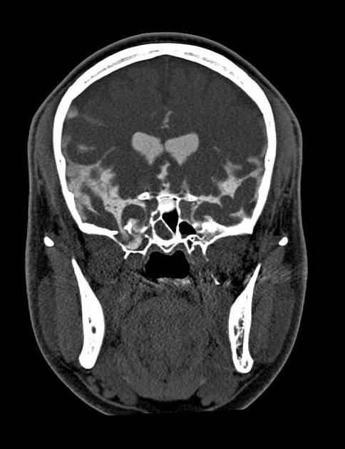
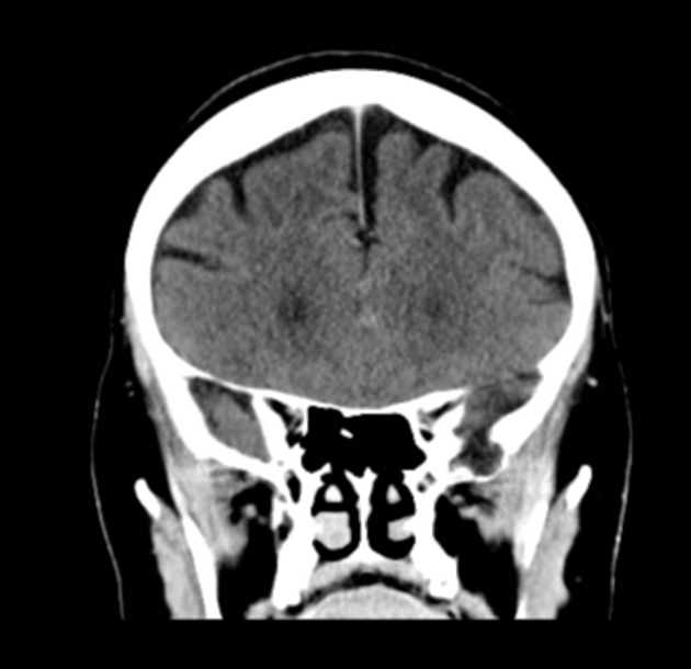
CT with thin bone algorithm images in multiple planes is essential in deliniating bony defects.
CT cisternography demonstrates sphenoid sinus pneumatization, a defect in the superior wall of the left lateral recess of the sphenoid sinus and anteromedial temporal lobe herniating through the bony defect.
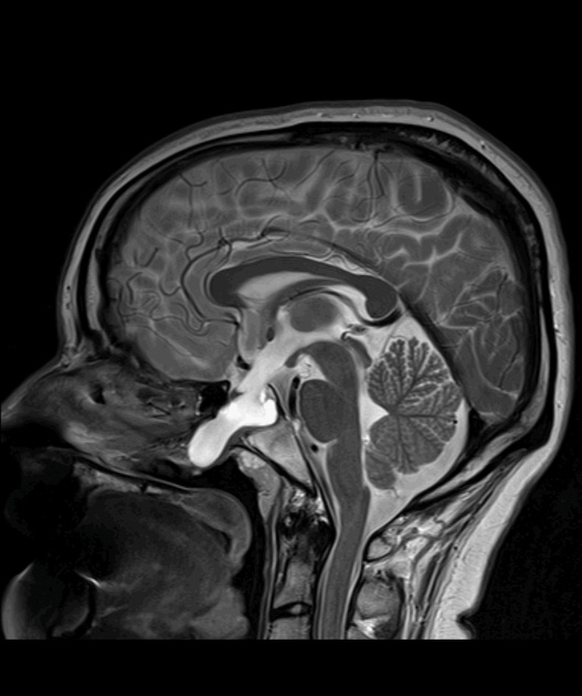
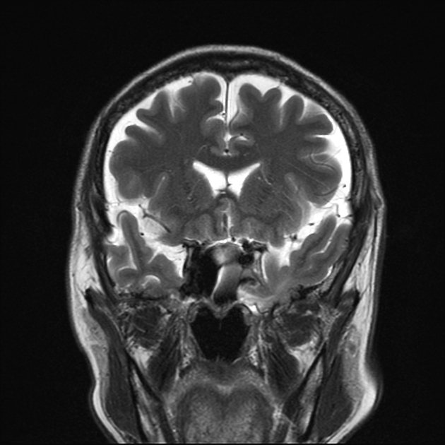
MRI
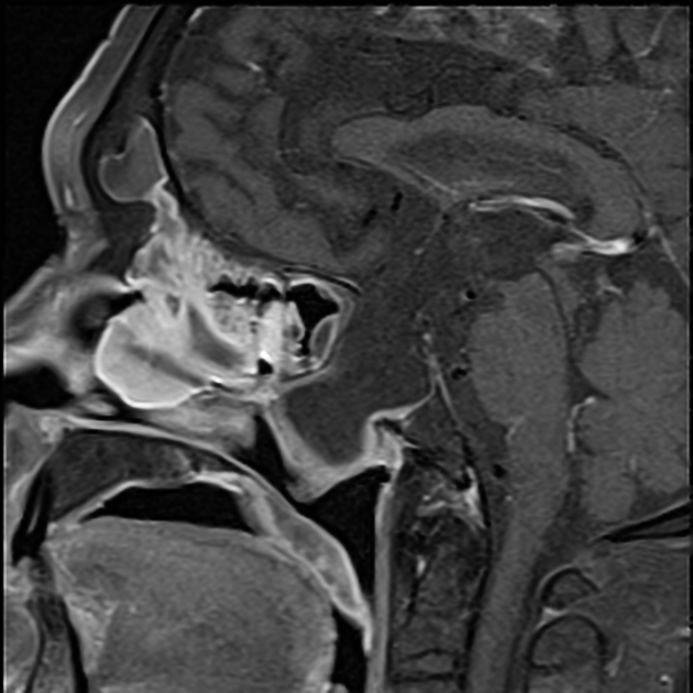
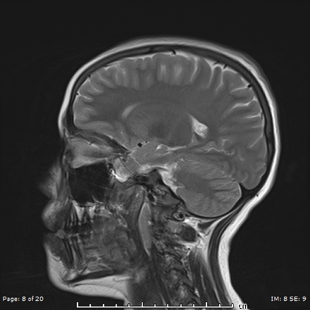
MRI is excellent at depicting the content of defects and on T2-weighted spin-echo sequence, the bright CSF signal is well appreciated against the signal void air of the paranasal sinuses 1.
MR cisternography is an alternative technique that can reveal CSF leakage in multiple planes. This can be performed with off-label use of intrathecal gadolinium and can be combined with iodinated contrast so that CT cisternography can be performed simultaneously 11.
Off-label warning: The use of gadolinium-based contrast agents for intrathecal administration is not approved by regulatory agencies such as the FDA. Read more: intrathecal gadolinium
Nuclear medicine
Radionuclide cisternography has been largely surplanted by CT and MRI 12.
Treatement and prognosis
The cornerstone of treatment of symptomatic sphenoid encephaloceles is surgical repair, usually endoscopically 7,8,10. If intracranial hypertension is present then it is essential that this is also actively managed to avoid recurrence.


 Unable to process the form. Check for errors and try again.