
This is a basic article for medical students and other non-radiologists
Investigating focal weakness makes up a large proportion of the workload for neurologists and neuroradiologists. A wide range of serious CNS disorders can present with focal weakness. Appropriate timely imaging can guide diagnosis and treatment.
Weakness can be due to changes in any part of the neurological system from muscle and neuromuscular junction, spinal cord or brain so history and examination are vital in guiding investigations and imaging.
On this page:
Images:
Reference article
This is a summary article; there is not a more in-depth reference article.
Summary
-
questions
is the weakness upper or lower motor neuron?
what is the distribution?
what was the speed of onset?
are there associated symptoms? e.g. headache, altered sensation
are there any co-morbidities? e.g. heart disease
have any other investigations been performed? e.g. lumbar puncture, nerve conduction studies/electromyography
-
investigations
-
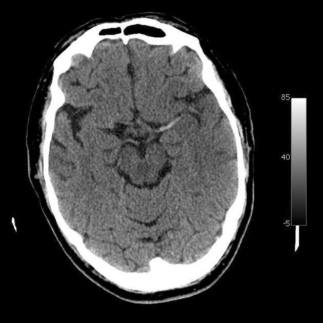
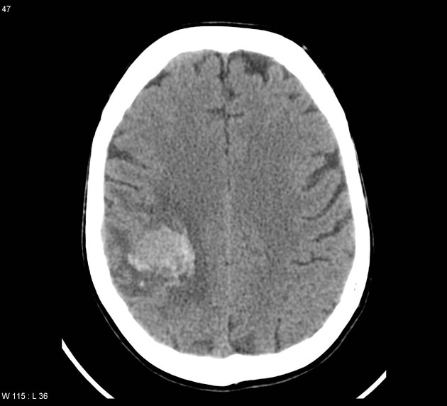
CT
most useful in the acute setting
readily available
able to exclude most hemorrhage and space occupying lesions
additional use of contrast may be performed
-
MRI
more sensitive than CT
improved contrast resolution
advanced imaging techniques help narrow the differential
lesion characterization
-
-
making the request
know the question you are trying to answer
if the patient may be for thrombolysis (in the setting of an acute ischemic stroke), know the time of symptom onset
be aware of the local imaging protocol
-
common pathology


 Unable to process the form. Check for errors and try again.