
Lateral cuneiform
Citation, DOI, disclosures and article data
At the time the article was created Craig Hacking had no recorded disclosures.
View Craig Hacking's current disclosuresAt the time the article was last revised Arlene Campos had no financial relationships to ineligible companies to disclose.
View Arlene Campos's current disclosures- Third cuneiform
- External cuneiform
- Cuneiforme lateral
- Lateral cuneiform bone
- Lateral cuneiform bones
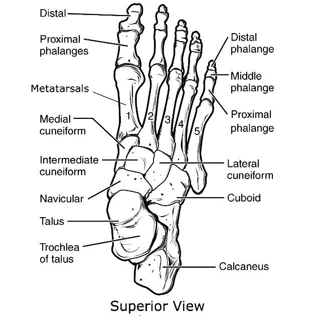
The lateral cuneiform is one of the tarsal bones located between the intermediate cuneiform and cuboid bones.
On this page:
Gross anatomy
Osteology
The lateral cuneiform is a wedge-shaped bone. It is smaller than the medial cuneiform and larger than the intermediate cuneiform. It lies edge downward, between the intermediate cuneiform and cuboid.
Articulations
anteriorly with the 2nd and 3rd metatarsals
posteriorly with the navicular
laterally with the cuboid
medially with the intermediate cuneiform
Attachments
Musculotendinous
flexor hallucis brevis: the proximal part of the lateral cuneiform undersurface gives rise to this muscle
tibialis posterior: one of its fibrous terminal tendon slips attaches to the narrow plantar surface
Ligamentous
plantar interosseous ligaments: arise from the lower part of the lateral cuneiform, forming part of the transverse arch of the foot, these are mainly cuneocuboid ligament and intercuneiform ligaments which help gliding and rotation in pedal pronation or supination and also when the forefoot is stressed, as in initial thrust of running and jumping
Relations
anterior: 2nd and 3rd metatarsal
posterior: navicular
lateral: cuboid
medial: intermediate cuneiform
Arterial supply
via the lateral tarsal artery, a continuing branch of the dorsalis pedis
Development
Ossification
The lateral cuneiform ossifies in the first year of life.
Related pathology
isolated lateral cuneiform fracture is rare, but has been documented in the literature 1
References
- 1. Shah K & Odgaard A. Fracture of the Lateral Cuneiform Only: A Rare Foot Injury. J Am Podiatr Med Assoc. 2007;97(6):483-5. doi:10.7547/0970483 - Pubmed
- 2. Susan Standring. Gray's Anatomy. (2008) ISBN: 9780443066849 - Google Books
Incoming Links
Related articles: Anatomy: Lower limb
- skeleton of the lower limb
- joints of the lower limb
-
hip joint
- ligaments
- muscles
- additional structures
- hip joint capsule
- zona orbicularis
- iliotibial band
-
hip bursae
- anterior
- iliopsoas bursa (iliopectineal bursa)
- lateral
- subgluteal bursae
- greater trochanteric bursa (subgluteus maximus bursa)
- subgluteus medius bursa
- subgluteus minimus bursa
- gluteofemoral bursa
- subgluteal bursae
- postero-inferior
- anterior
- ossification centers
-
knee joint
- ligaments
- anterior cruciate ligament
- posterior cruciate ligament
- medial collateral ligament
- lateral collateral ligament
- meniscofemoral ligament (mnemonic)
-
posterolateral ligamentous complex
- arcuate ligament
- patellar tendon and quadriceps tendon
- anterolateral ligament
- posterior oblique ligament
- oblique popliteal ligament
- medial patellofemoral ligament
- additional structures
- extensor mechanism of the knee
- groove for the popliteus tendon
- knee bursae
- anterior bursae
- medial bursae
- lateral bursae
- posterior bursae
- knee capsule
- lateral patellar retinaculum
- medial patellar retinaculum
- menisci
- pes anserinus (mnemonic)
- ossification centers
- ligaments
- tibiofibular joints
-
ankle joint
- regional anatomy
- medial ankle
- lateral ankle
- anterior ankle
- ligaments
- medial collateral (deltoid) ligament
- lateral collateral ligament
- additional structures
- ankle bursae
- ossification centers of the ankle
- variants
- regional anatomy
- foot joints
- subtalar joint
- mid-tarsal (Chopart) joint
-
tarsometatarsal (Lisfranc) joint
- ligaments
- intermetatarsal joint
- metatarsophalangeal joint
- interphalangeal joint
- ossification centers
-
hip joint
- spaces of the lower limb
-
muscles of the lower limb
- muscles of the pelvic group
- muscles of the thigh
- muscles of the leg
- anterior compartment of the leg
- posterior compartments of the leg
- lateral compartment of the leg
- muscles of the foot
- dorsal muscles
- plantar muscles
- 1st layer
- 2nd layer
- 3rd layer
- 4th layer
- accessory muscles of the lower limb
- accessory gluteal muscles
-
accessory muscles of the ankle
- accessory peroneal muscles
- accessory flexor digitorum longus muscle
- accessory soleus muscle
- peroneocalcaneus internus muscle
- tibiocalcaneus internus muscle
- extensor hallucis capsularis tendon
- anterior fibulocalcaneus muscle
- accessory extensor digiti secundus muscle
- tibioastragalus anticus of Gruber muscle
- vascular supply of the lower limb
- arterial supply of the lower limb
- venous drainage of the lower limb
- innervation of the lower limb
- lymphatic system of the lower limb
- lymphatic pathways
- anteromedial group
- anterolateral group
- posteromedial group
- posterolateral group
- lower limb lymph nodes
- lymphatic pathways


 Unable to process the form. Check for errors and try again.