Left atrium
Updates to Article Attributes
The left atrium (LA) (plural: atria) is one of the four chambers of the heart. It receives oxygenated blood from the pulmonary circulation that is then delivered to the left ventricle (LV) and then into the systemic circulation.
Gross anatomy
The left atrium is grossly cuboidal,. It is the most superior and posterior portion of the heart and is obscured anteriorly by the pulmonary trunk and ascending aorta. It lies mostly to the right of the left ventricle.
The left atrium is separated from the right atrium by the intertribal (interatrial)interatrial septum, with thea small depression, the fossa ovaleovalis, at the site of the closed foramen ovale. It is smaller by volume than the right atrium but with thicker walls.
The left atrial appendage is long and narrow, curving anteriorly from the left around the pulmonary trunk, overlying part of the left coronary artery. Its inner surface is lined by the small pectinate muscles, unlike the main cavity which has smooth walls.
Blood enters on either side via four pulmonary veins, the left and right superior and inferior, and exits via the left atrioventricular opening through the mitral valve (MV), into the left ventricle. The mitral valve is bicuspid with anterior and posterior leaflets supported by the left atrium.
Arterial supply
left circumflex artery: a branch of the left coronary artery that feeds small atrial branches
Venous drainage
great cardiac vein: drains into the left side of the coronary sinus, and on into the right atrium
oblique vein of Marshall: drains into the coronary sinus and is not present in all patients
tiny myocardial thebesian veins drain directly into the right ventricle
Innervation
Variant anatomy
accessory left atrial appendage: common variant in which small diverticular structure is seen on the superior right side
left atrial diverticulum: a pouch-like diverticulum, differentiated from accessory atrial appendage by this smooth shape suggesting a lack of pectinate muscles
coumadin ridge: band-like embryological remnant between the left superior pulmonary vein and the left atrial appendage
Radiographic features
On contrast-enhanced chest CT and cardiac MRI, the left atrium when measured on axial slices can be considered enlarged when the anteroposterior diameter is ≥50 mm (male) and ≥45 mm (female) 6.
Development
In the fetal heart, the left and right atria communicate via the foramen ovale in the interatrial septum. Both atria develop from a single primitive atrium. The only remnant of this in the left atrium is the atrial appendage. The smooth-walled main cavity of the left atrium develops from the pulmonary veins. See development of the heart.
History and etymology
"Atrium" is the Latin word for "court"an entrance hall, referring to the central area in a Roman house from which one could enter various chambers. It was entered through the "ostium" of a Roman house.
Practical points
The interatrial septum may be difficult to identify on CT. This is especially true of the fossa ovalis, being even thinner, which may be unidentifiable and mistaken as an atrial septal defect.
The junction of the left atrial appendage and left superior pulmonary vein forms a muscular ridge. This ridge varies in size and may be large and pronounced, and thus mistaken for a pedunculated mass or thrombus in the left lateral wall.
The left atrium lies directly in front of the oesophagus and is, therefore, an appropriate window for transoesophageal echocardiography.
Related pathology
-<p>The <strong>left atrium </strong>(<strong>LA</strong>) (plural: atria) is one of the four chambers of the <a href="/articles/heart">heart</a>. It receives oxygenated blood from the pulmonary circulation that is then delivered to the <a href="/articles/left-ventricle">left ventricle (LV)</a> and then into the systemic circulation.</p><h4>Gross anatomy</h4><p>The left atrium is grossly cuboidal,. It is the most superior and posterior portion of the heart and is obscured anteriorly by the <a href="/articles/pulmonary-trunk">pulmonary trunk</a> and <a href="/articles/ascending-aorta">ascending aorta</a>. It lies mostly to the right of the left ventricle.</p><p>The left atrium is separated from the right atrium by the <a href="/articles/atrial-septum-1">intertribal (interatrial) septum</a>, with the small depression, the <a href="/articles/fossa-ovale">fossa ovale</a>, at the site of the closed <a href="/articles/foramen-ovale-cardiac-1">foramen ovale</a>. It is smaller by volume than the right atrium but with thicker walls.</p><p>The <a href="/articles/left-atrial-appendage">left atrial appendage</a> is long and narrow, curving anteriorly from the left around the pulmonary trunk, overlying part of the left coronary artery. Its inner surface is lined by the small pectinate muscles, unlike the main cavity which has smooth walls.</p><p>Blood enters on either side via four <a href="/articles/pulmonary-veins">pulmonary veins</a>, the left and right superior and inferior, and exits via the left atrioventricular opening through the <a href="/articles/mitral-valve">mitral valve (MV)</a>, into the left ventricle. The mitral valve is bicuspid with anterior and posterior leaflets supported by the left atrium.</p><h4>Arterial supply</h4><ul><li>-<a href="/articles/circumflex-artery">left circumflex artery</a>: a branch of the <a href="/articles/left-main-coronary-artery-1">left coronary artery</a> that feeds small atrial branches</li></ul><h4>Venous drainage</h4><ul>-<li>-<a href="/articles/great-cardiac-vein">great cardiac vein</a>: drains into the left side of the <a href="/articles/coronary-sinus">coronary sinus</a>, and on into the right atrium</li>-<li>-<a href="/articles/vein-of-marshall-1">oblique vein of Marshall</a>: drains into the coronary sinus and is not present in all patients</li>-<li>tiny myocardial <a href="/articles/smallest-cardiac-veins">thebesian veins</a> drain directly into the right ventricle</li>- +<p>The <strong>left atrium </strong>(<strong>LA</strong>) (plural: atria) is one of the four chambers of the <a href="/articles/heart">heart</a>. It receives oxygenated blood from the pulmonary circulation that is then delivered to the <a href="/articles/left-ventricle">left ventricle</a> and then into the systemic circulation.</p><h4>Gross anatomy</h4><p>The left atrium is grossly cuboidal. It is the most superior and posterior portion of the heart and is obscured anteriorly by the <a href="/articles/pulmonary-trunk">pulmonary trunk</a> and <a href="/articles/ascending-aorta">ascending aorta</a>. It lies mostly to the right of the left ventricle.</p><p>The left atrium is separated from the right atrium by the <a href="/articles/atrial-septum-1">interatrial septum</a>, with a small depression, the <a href="/articles/fossa-ovalis">fossa ovalis</a>, at the site of the closed <a href="/articles/foramen-ovale-cardiac-1">foramen ovale</a>. It is smaller by volume than the right atrium but with thicker walls.</p><p>The <a href="/articles/left-atrial-appendage">left atrial appendage</a> is long and narrow, curving anteriorly from the left around the pulmonary trunk, overlying part of the left coronary artery. Its inner surface is lined by the small pectinate muscles, unlike the main cavity which has smooth walls.</p><p>Blood enters on either side via four <a href="/articles/pulmonary-veins">pulmonary veins</a>, the left and right superior and inferior, and exits via the left atrioventricular opening through the <a href="/articles/mitral-valve">mitral valve</a>, into the left ventricle. The mitral valve is bicuspid with anterior and posterior leaflets supported by the left atrium.</p><h4>Arterial supply</h4><ul><li><p><a href="/articles/circumflex-artery">left circumflex artery</a>: a branch of the <a href="/articles/left-main-coronary-artery-1">left coronary artery</a> that feeds small atrial branches</p></li></ul><h4>Venous drainage</h4><ul>
- +<li><p><a href="/articles/great-cardiac-vein">great cardiac vein</a>: drains into the left side of the <a href="/articles/coronary-sinus">coronary sinus</a>, and on into the right atrium</p></li>
- +<li><p><a href="/articles/vein-of-marshall-1">oblique vein of Marshall</a>: drains into the coronary sinus and is not present in all patients</p></li>
- +<li><p>tiny myocardial <a href="/articles/smallest-cardiac-veins">thebesian veins</a> drain directly into the right ventricle</p></li>
-<li>-<a href="/articles/accessory-left-atrial-appendage">accessory left atrial appendage</a>: common variant in which small diverticular structure is seen on the superior right side</li>-<li>-<a href="/articles/left-atrial-diverticulum">left atrial diverticulum</a>: a pouch-like diverticulum, differentiated from accessory atrial appendage by this smooth shape suggesting a lack of pectinate muscles</li>-<li>-<a href="/articles/coumadin-ridge">coumadin ridge</a>: band-like embryological remnant between the left superior pulmonary vein and the left atrial appendage</li>-</ul><h4>Radiographic features</h4><p>On contrast-enhanced chest CT and cardiac MRI, the left atrium when measured on axial slices can be considered enlarged when the anteroposterior diameter is ≥50 mm (male) and ≥45 mm (female) <sup>6</sup>.</p><h4>Development</h4><p>In the fetal heart, the left and right atria communicate via the <a href="/articles/foramen-ovale-cardiac-1">foramen ovale</a> in the interatrial septum. Both atria develop from a single primitive atrium. The only remnant of this in the left atrium is the atrial appendage. The smooth-walled main cavity of the left atrium develops from the pulmonary veins. See <a href="/articles/development-of-the-heart-1">development of the heart</a>.</p><h4>History and etymology</h4><p>"Atrium" is the Latin word for "court", referring to the central area in a Roman house from which one could enter various chambers. It was entered through the "ostium" of a Roman house.</p><h4>Practical points</h4><p>The interatrial septum may be difficult to identify on CT. This is especially true of the fossa ovalis, being even thinner, which may be unidentifiable and mistaken as an atrial septal defect.</p><p>The junction of the left atrial appendage and left superior pulmonary vein forms a muscular ridge. This ridge varies in size and may be large and pronounced, and thus mistaken for a pedunculated mass or thrombus in the left lateral wall.</p><p>The left atrium lies directly in front of the oesophagus and is, therefore, an appropriate window for transoesophageal echocardiography.</p><h4>Related pathology</h4><ul>-<li><a href="/articles/atrial-septal-defect-2">atrial septal defect (ASD)</a></li>-<li><a href="/articles/interatrial-septal-aneurysm">interatrial septal aneurysm</a></li>-<li><a href="/articles/left-atrial-enlargement">left atrial enlargement</a></li>-<li><a href="/articles/mitral-valve-regurgitation">mitral regurgitation</a></li>-<li><a href="/articles/mitral-valve-stenosis-1">mitral stenosis</a></li>-<li>-<a href="/articles/porcelain-left-atrium">porcelain left atrium</a> a.k.a. coconut left atrium</li>- +<li><p><a href="/articles/accessory-left-atrial-appendage">accessory left atrial appendage</a>: common variant in which small diverticular structure is seen on the superior right side</p></li>
- +<li><p><a href="/articles/left-atrial-diverticulum">left atrial diverticulum</a>: a pouch-like diverticulum, differentiated from accessory atrial appendage by this smooth shape suggesting a lack of pectinate muscles</p></li>
- +<li><p><a href="/articles/coumadin-ridge">coumadin ridge</a>: band-like embryological remnant between the left superior pulmonary vein and the left atrial appendage</p></li>
- +</ul><h4>Radiographic features</h4><p>On contrast-enhanced chest CT and cardiac MRI, the left atrium when measured on axial slices can be considered enlarged when the anteroposterior diameter is ≥50 mm (male) and ≥45 mm (female) <sup>6</sup>.</p><h4>Development</h4><p>In the fetal heart, the left and right atria communicate via the <a href="/articles/foramen-ovale-cardiac-1">foramen ovale</a> in the interatrial septum. Both atria develop from a single primitive atrium. The only remnant of this in the left atrium is the atrial appendage. The smooth-walled main cavity of the left atrium develops from the pulmonary veins. See <a href="/articles/development-of-the-heart-1">development of the heart</a>.</p><h4>History and etymology</h4><p>"Atrium" is the Latin word for an entrance hall, referring to the central area in a Roman house from which one could enter various chambers. It was entered through the "ostium" of a Roman house.</p><h4>Practical points</h4><p>The interatrial septum may be difficult to identify on CT. This is especially true of the fossa ovalis, being even thinner, which may be unidentifiable and mistaken as an atrial septal defect.</p><p>The junction of the left atrial appendage and left superior pulmonary vein forms a muscular ridge. This ridge varies in size and may be large and pronounced, and thus mistaken for a pedunculated mass or thrombus in the left lateral wall.</p><p>The left atrium lies directly in front of the oesophagus and is, therefore, an appropriate window for transoesophageal echocardiography.</p><h4>Related pathology</h4><ul>
- +<li><p><a href="/articles/atrial-septal-defect-2">atrial septal defect (ASD)</a></p></li>
- +<li><p><a href="/articles/interatrial-septal-aneurysm">interatrial septal aneurysm</a></p></li>
- +<li><p><a href="/articles/left-atrial-enlargement">left atrial enlargement</a></p></li>
- +<li><p><a href="/articles/mitral-valve-regurgitation">mitral regurgitation</a></p></li>
- +<li><p><a href="/articles/mitral-valve-stenosis-1">mitral stenosis</a></p></li>
- +<li><p><a href="/articles/porcelain-left-atrium">porcelain left atrium</a> a.k.a. coconut left atrium</p></li>
References changed:
- 1. Vincent B. Ho, Gautham P. Reddy. Cardiovascular Imaging. (2010) ISBN: 9781416053354 - <a href="http://books.google.com/books?vid=ISBN9781416053354">Google Books</a>
- 2. Broderick L, Brooks G, Kuhlman J. Anatomic Pitfalls of the Heart and Pericardium. Radiographics. 2005;25(2):441-53. <a href="https://doi.org/10.1148/rg.252045075">doi:10.1148/rg.252045075</a> - <a href="https://www.ncbi.nlm.nih.gov/pubmed/15798062">Pubmed</a>
- 3. Abbara S, Mundo-Sagardia J, Hoffmann U, Cury R. Cardiac CT Assessment of Left Atrial Accessory Appendages and Diverticula. AJR Am J Roentgenol. 2009;193(3):807-12. <a href="https://doi.org/10.2214/AJR.08.2229">doi:10.2214/AJR.08.2229</a> - <a href="https://www.ncbi.nlm.nih.gov/pubmed/19696296">Pubmed</a>
- 4. Chummy S. Sinnatamby. Last's Anatomy. (2011) ISBN: 9780702033957 - <a href="http://books.google.com/books?vid=ISBN9780702033957">Google Books</a>
- 5. Keith L. Moore, T. V. N. Persaud, Mark G. Torchia. The Developing Human. (2015) ISBN: 9780323313384 - <a href="http://books.google.com/books?vid=ISBN9780323313384">Google Books</a>
- 6. Eifer D, Nguyen E, Thavendiranathan P, Hanneman K. Diagnostic Accuracy of Sex-Specific Chest CT Measurements Compared With Cardiac MRI Findings in the Assessment of Cardiac Chamber Enlargement. AJR Am J Roentgenol. 2018;211(5):993-9. <a href="https://doi.org/10.2214/AJR.18.19805">doi:10.2214/AJR.18.19805</a> - <a href="https://www.ncbi.nlm.nih.gov/pubmed/30240288">Pubmed</a>
- 1. Ho V, Reddy PR. Cardiovascular Imaging, 2-Volume Set. Saunders. ISBN:1416053352. <a href="http://books.google.com/books?vid=ISBN1416053352">Read it at Google Books</a> - <a href="http://www.amazon.com/gp/product/1416053352">Find it at Amazon</a><span class="auto"></span>
- 2. Broderick LS, Brooks GN, Kuhlman JE. Anatomic pitfalls of the heart and pericardium. Radiographics. 2005;25 (2): 441-53. <a href="http://dx.doi.org/10.1148/rg.252045075">doi:10.1148/rg.252045075</a> - <a href="http://www.ncbi.nlm.nih.gov/pubmed/15798062">Pubmed citation</a><span class="auto"></span>
- 3. Abbara S, Mundo-Sagardia JA, Hoffmann U et-al. Cardiac CT assessment of left atrial accessory appendages and diverticula. AJR Am J Roentgenol. 2009;193 (3): 807-12. <a href="http://dx.doi.org/10.2214/AJR.08.2229">doi:10.2214/AJR.08.2229</a> - <a href="http://www.ncbi.nlm.nih.gov/pubmed/19696296">Pubmed citation</a><span class="auto"></span>
- 4. Sinnatamby CS. Last's Anatomy. Churchill Livingstone. (2011) ISBN:0702033952. <a href="http://books.google.com/books?vid=ISBN0702033952">Read it at Google Books</a> - <a href="http://www.amazon.com/gp/product/0702033952">Find it at Amazon</a><span class="ref_v3"></span>
- 5. Moore KL, Persuad TVN, Torchia MG. The Developing Human: Clinically Oriented Embryology, 10e. Saunders. ISBN:0323313388. <a href="http://books.google.com/books?vid=ISBN0323313388">Read it at Google Books</a> - <a href="http://www.amazon.com/gp/product/0323313388">Find it at Amazon</a><span class="auto"></span>
- 6. Eifer DA, Nguyen ET, Thavendiranathan P, Hanneman K. Diagnostic Accuracy of Sex-Specific Chest CT Measurements Compared With Cardiac MRI Findings in the Assessment of Cardiac Chamber Enlargement. (2018) AJR. American journal of roentgenology. 211 (5): 993-999. <a href="https://doi.org/10.2214/AJR.18.19805">doi:10.2214/AJR.18.19805</a> - <a href="https://www.ncbi.nlm.nih.gov/pubmed/30240288">Pubmed</a> <span class="ref_v4"></span>
Image 1 Annotated image (Axial C+ arterial phase) ( create )

Image 2 Annotated image (Axial C+ coronary angiogram) ( create )

Image 3 Diagram ( update )

Image 4 Annotated image (Coronal C+ arterial phase) ( create )

Image 5 Annotated image (Sagittal C+ arterial phase) ( create )

Image 6 Diagram ( update )
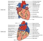
Image 7 Diagram ( update )
