
Historically, the lymph nodes in the neck have been anatomically divided into at least six neck lymph node levels for head and neck cancer staging and therapy-planning purposes. Differing definitions exist across specialties 1-4. The following is a synthesis of radiologically useful boundaries for each level.
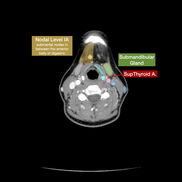
Level I: submental and submandibular
superiorly: mylohyoid muscle and mandible
inferiorly: inferior border of the hyoid bone
anteriorly: platysma muscle
posteriorly: posterior border of the submandibular gland
There are two sublevels:
level Ia (submental nodes): anteromedial between the anterior bellies of both digastric muscles
level Ib (submandibular nodes): posterolateral to the anterior belly of the digastric muscles
Level II: upper internal jugular (deep cervical) chain
superiorly: base of the skull at the jugular fossa
inferiorly: inferior border of the hyoid bone
anteriorly: posterior border of the submandibular gland
posterolaterally: posterior border of the sternocleidomastoid muscle
medially: medial border of the internal carotid artery
There are two sublevels:
level IIa: inseparable from or anterior to the posterior edge of the internal jugular vein; includes jugulodigastric nodal group
level IIb: posterior to and separable by a fat plane from the internal jugular vein
Level III: middle internal jugular (deep cervical) chain
superiorly: inferior border of the hyoid bone
inferiorly: inferior border of the cricoid cartilage
anteriorly: anterior border of the sternocleidomastoid muscle
posterolaterally: posterior border of the sternocleidomastoid muscle
medially: medial border of the common carotid artery
Level IV: lower internal jugular (deep cervical) chain
superiorly: inferior border of the cricoid cartilage
inferiorly: level of the clavicle
anteriorly: anterior border of the sternocleidomastoid muscle
posterolaterally: oblique line drawn through the posterolateral edge of the sternocleidomastoid muscle and the lateral edge of the anterior scalene muscle 2
medially: medial border of the common carotid artery
includes medial supraclavicular nodes including Virchow node 1
Level V: posterior triangle
superiorly: skull base at the apex of the convergence of sternocleidomastoid and trapezius muscles
inferiorly: level of the clavicle
anteromedially: posterior border of the sternocleidomastoid muscle
posterolaterally: anterior border of the trapezius muscle
There are two sublevels:
level Va: superior half, superior to inferior border of the cricoid cartilage (posterior to levels II and III); includes spinal accessory nerve
level Vb: inferior half, inferior to inferior border of the cricoid cartilage (posterior to level IV); includes lateral supraclavicular nodes 1
Level VI: central (anterior) compartment
superiorly: inferior border of hyoid bone
inferiorly: superior border of manubrium (suprasternal notch)
anteriorly: platysma muscle 8
posteriorly: trachea (medially) and prevertebral space (laterally) 8
laterally: medial borders of both common carotid arteries (medial to levels III and IV)
includes anterior jugular, pretracheal, paratracheal, prelaryngeal/precricoid (Delphian), and perithyroidal nodes
Termination
All the levels above eventually drain to the jugular trunk of their respective side and then to the right lymphatic duct or the thoracic duct (left) 10.
Variant anatomic boundaries
Minor variations to the above anatomic boundaries have been described.
For example, head and neck surgeons may use different intraoperative landmarks 1,4:
levels IIa and IIb are separated by the vertical plane defined by the spinal accessory nerve
the medial border of levels III and IV and lateral border of level VI is defined by the lateral border of the sternohyoid muscle
the posterior border of levels II through IV and anterior border of level V may also be defined by the plane of sensory branches of the cervical plexus
In addition, radiologists may describe supraclavicular nodes separately from levels IV and V using the transverse level of the clavicle as the border 9.
Additional neck levels
The above classification is not inclusive of several important nodal groups in the head and neck:
Other classification systems include some of these regions, but a consensus approach has not been reached. For example, with respect to "level VII," radiologists may apply this label to superior mediastinal nodes 2, radiation oncologists may apply this label to "prevertebral nodes" 3, and head and neck surgeons omit this level from their nomenclature altogether 1. Thus, it is best to name lymph node groups outside of the established levels I-VI. If "level VII" is used for superior mediastinal lymph nodes, it should refer to the extension of the paratracheal chain below the suprasternal notch but above the level of the brachiocephalic artery 4.


 Unable to process the form. Check for errors and try again.