
The olfactory nerve is the first (CN I) cranial nerve (TA: nervus olfactorius or nervus cranialis I) and is responsible for conveying the sense of smell from the nasal cavity to the brain. Strictly speaking, the term olfactory 'nerve' refers only to the short first order neurones (olfactory filaments) located on the olfactory mucosa. The olfactory bulb, tract and striae are often erroneously referred together as the olfactory nerve, but they are actually an extension of the central nervous system (see olfactory system).
On this page:
Summary
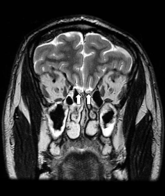
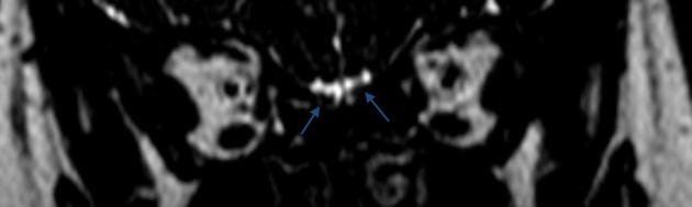
location: olfactory groove; anterior cranial fossa near the midline
branches and supply: 20 or so olfactory filaments in the roof of the nasal cavity
Gross anatomy
Course
Olfactory filaments
The bipolar cell (olfactory sensory neurone, OSN) is the first-order sensory neurone located within the specialised olfactory neuroepithelium, within the 'olfactory cleft' (bounded superiorly by the cribriform plate of the ethmoid bone, medially by the septum and laterally by the superior turbinate) of the nasal cavity. This cell is analogous to the sensory cells of spinal nerves, whose cell bodies reside in the dorsal root ganglion. There are thought to be approximately 6-30 million OSN in humans. Their central processes (axons) form filaments which pass through the cribriform plate, pierce the dura mater and relay in the olfactory bulb. Together, these filaments form the 'olfactory nerve'.
Olfactory bulb
The olfactory bulbs are paired structures that forms the first synaptic connection with the olfactory fossa. It houses the cell bodies of mitral cells, the second-order neurones of the olfactory system.
Olfactory tract
The central process of these second-order neurones forms the olfactory tract which courses posteriorly, superior to the olfactory groove of the anterior cranial fossa, and inferior to the olfactory sulcus (lateral to gyrus rectus, and medial to the orbital gyri).
Olfactory striae
Anterior to the anterior perforated substance, the olfactory tract divides to form the medial and lateral olfactory striae. This triangular area of division is referred to as the olfactory trigone. The lateral olfactory striae project to the uncus. The medial olfactory striae ultimately project to the hypothalamus and brainstem nuclei.
Arterial supply
The sphenopalatine, anterior, and posterior ethmoid arteries supply the olfactory neurones and mucosa. The olfactory bulb, tract, and straie are supplied by the olfactory artery, a branch of the anterior cerebral artery 6.
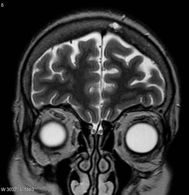
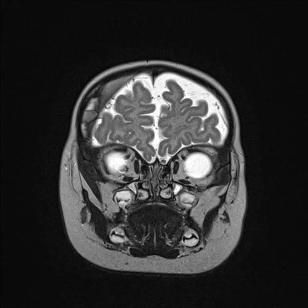
Radiographic features
MRI
Coronal images are the best to depict the olfactory nerve as it is situated deep in the olfactory groove.


 Unable to process the form. Check for errors and try again.