
Parahippocampal gyrus
Last revised by Zander Hendrik Engelbrecht
on 1 Mar 2022
Citation, DOI, disclosures and article data
Citation:
Goel A, Engelbrecht Z, Gan C, et al. Parahippocampal gyrus. Reference article, Radiopaedia.org (Accessed on 17 Mar 2025) https://doi.org/10.53347/rID-34427
rID:
34427
Article created:
19 Feb 2015,
Ayush Goel
Disclosures:
At the time the article was created Ayush Goel had no recorded disclosures.
View Ayush Goel's current disclosures
Last revised:
1 Mar 2022,
Zander Hendrik Engelbrecht
Disclosures:
At the time the article was last revised Zander Hendrik Engelbrecht had no recorded disclosures.
View Zander Hendrik Engelbrecht's current disclosures
Revisions:
6 times, by
5 contributors -
see full revision history and disclosures
Systems:
Sections:
Synonyms:
- Parahippocampal gyri
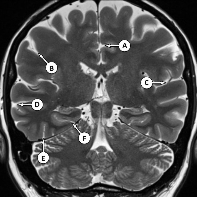
The parahippocampal gyrus is a cortical ridge in the medial temporal lobe, located superior to the tentorium. Anteriorly it curves to form the uncus of the temporal lobe and posteriorly it appears continuous with the lingual gyrus.
It covers the the hippocampus medially and amygdala anteromedially. Inferolaterally it is bounded by the collateral sulcus.
Quiz questions
References
- 1. Blaizot X, Martinez-Marcos A, Arroyo-Jimenez Md Mdel M et-al. The parahippocampal gyrus in the baboon: anatomical, cytoarchitectonic and magnetic resonance imaging (MRI) studies. Cereb. Cortex. 2004;14 (3): 231-46. Pubmed citation
- 2. Last RJ, McMinn RMH. Last's Anatomy Regional and Applied. 9th ed. New York: Churchill Livingstone; 2003. p. 581.
Incoming Links
Articles:
- Entorhinal cortical atrophy score
- Head ultrasound
- Limbic lobe
- Fusiform gyrus
- Basal vein of Rosenthal
- Entorhinal cortex
- Lingual gyrus
- Limbic system
- Temporal lobe
- Hippocampus
- Mesial temporal lobe
- Posterior choroidal artery infarct
- Papez circuit
- Uncus
- Olfactory system
- Medial occipitotemporal gyrus
- Subiculum
- Collateral sulcus
- Mesial temporal sclerosis
Multiple choice questions:
Related articles: Anatomy: Brain
-
brain
- grey matter
- white matter
-
cerebrum
-
cerebral hemisphere (telencephalon)
- cerebral lobes and gyri
- frontal lobe
- parietal lobe
-
occipital lobe
- occipital pole
- lingual gyrus
- fusiform gyrus (Brodmann area 37)
- calcarine (visual) cortex
- cuneus
- temporal lobe
- basal forebrain
- limbic system
- insula
-
cerebral sulci and fissures (A-Z)
- calcarine fissure
- callosal sulcus
- central (Rolandic) sulcus
- cingulate sulcus
- collateral sulcus
- inferior frontal sulcus
- inferior occipital sulcus
- inferior temporal sulcus
- interhemispheric fissure
- intraparietal sulcus
- lateral (Sylvian) sulcus
- lateral occipital sulcus
- marginal sulcus
- occipitotemporal sulcus
- olfactory sulcus
- paracentral sulcus
- paraolfactory sulcus
- parieto-occipital fissure
- posterior parolfactory sulcus
- precentral sulcus
- preoccipital notch
- postcentral sulcus
- rhinal sulcus
- rostral sulcus
- subparietal sulcus
- superior frontal sulcus
- superior occipital sulcus
- superior temporal sulcus
- cortical histology
- cerebral lobes and gyri
- white matter tracts
- deep grey matter
-
pituitary gland
- posterior pituitary and stalk (part of diencephalon)
- anterior pituitary
- inferior hypophyseal arterial circle
- diencephalon
-
cerebral hemisphere (telencephalon)
-
brainstem
- midbrain (mesencephalon)
- pons (part of metencephalon)
- medulla oblongata (myelencephalon)
- white matter
-
grey matter
- non-cranial nerve
-
cranial nerve nuclei
- oculomotor nucleus
- Edinger-Westphal nucleus
- trochlear nucleus
- motor nucleus of CN V
- mesencephalic nucleus of CN V
- main sensory nucleus of CN V
- spinal nucleus of CN V
- abducent nucleus
- facial nucleus
- superior salivatory nucleus
- cochlear nuclei
- vestibular nuclei
- inferior salivatory nucleus
- solitary tract nucleus
- ambiguus nucleus
- dorsal vagal motor nucleus
- hypoglossal nucleus
-
cerebellum (part of metencephalon)
- vermis
- cerebellar hemisphere
- cerebellar peduncles
- cranial meninges (meninx primitiva)
- CSF spaces
-
cranial nerves (mnemonic)
- olfactory nerve (CN I)
- optic nerve (CN II)
- oculomotor nerve (CN III)
- trochlear nerve (CN IV)
- trigeminal nerve (CN V) (mnemonic)
- abducens nerve (CN VI)
- facial nerve (CN VII) (segments mnemonic | branches mnemonic)
-
vestibulocochlear nerve (CN VIII)
- vestibular ganglion (Scarpa's ganglion)
- glossopharyngeal nerve (CN IX)
- vagus nerve (CN X)
- spinal accessory nerve (CN XI)
- hypoglossal nerve (CN XII)
- functional neuroanatomy
- CNS development
- cerebral vascular supply
- arteries
- vascular territories
-
circle of Willis
- internal carotid artery (ICA) (segments)
- vertebral artery
-
normal variants
- intracranial arterial fenestration
- internal carotid artery (ICA)
- anterior cerebral artery (ACA)
- middle cerebral artery (MCA)
- posterior cerebral artery (PCA)
- basilar artery
- persistent carotid-vertebrobasilar artery anastomoses (mnemonic)
- vertebral artery
- ophthalmic artery
-
cerebral venous system
-
dural venous sinuses
- basilar venous plexus
- cavernous sinus (mnemonic)
- clival diploic veins
- inferior petro-occipital vein
- inferior petrosal sinus
- inferior sagittal sinus
- intercavernous sinus
- internal carotid artery venous plexus of Rektorzik
- jugular bulb
- marginal sinus
- occipital sinus
- sigmoid sinus
- sphenoparietal sinus
- straight sinus
- superior petrosal sinus
- superior sagittal sinus
- torcula herophili
- transverse sinus
-
cerebral veins
-
superficial veins of the brain
- superior cerebral veins (superficial cerebral veins)
- inferior cerebral veins
- superficial middle cerebral vein
- superior anastomotic vein (of Trolard)
- inferior anastomotic vein (of Labbe)
-
superficial veins of the brain
-
deep veins of the brain
- great cerebral vein (of Galen)
- venous circle of Trolard
- normal variants
-
dural venous sinuses
- arteries
- glymphatic pathway


 Unable to process the form. Check for errors and try again.