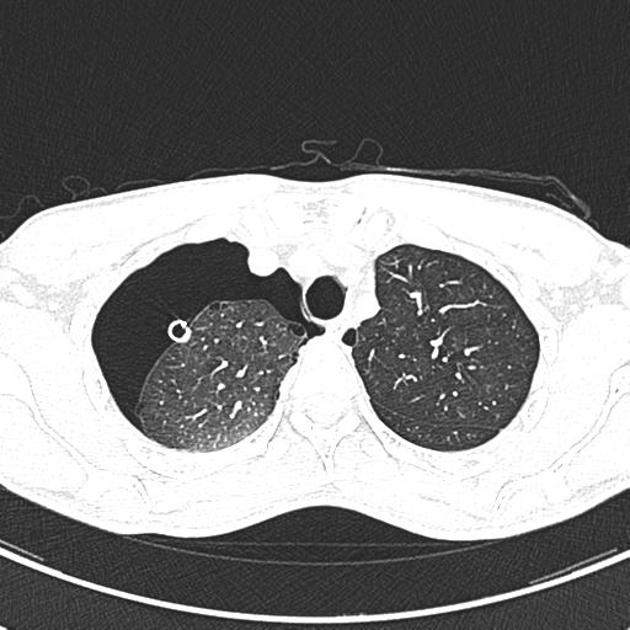
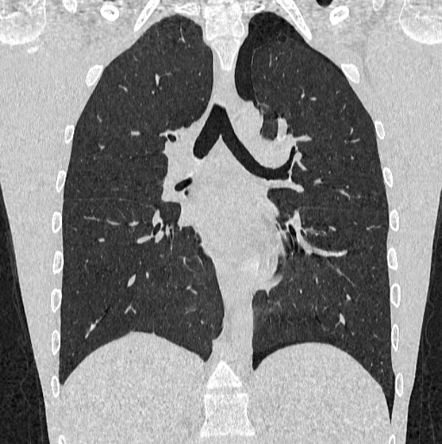
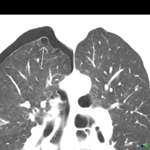
Pulmonary blebs are small subpleural air-filled circumscribed cystic spaces, usually less than 1 cm in diameter 4. There is an association with small airways disease. Rupture causes pneumothorax and is associated with tobacco smoking.
On this page:
Epidemiology
Blebs are a very common finding in otherwise normal individuals. They are often found in young patients. They are more common in thin patients and in cigarette smokers 1.
Clinical presentation
In the vast majority of cases, blebs remain asymptomatic. Occasionally they are thought to rupture resulting in a pneumothorax.
Pathology
Blebs are thought to occur as a result of subpleural alveolar rupture, due to strain overload of the elastic fibers.
Pulmonary bullae are, like blebs, cystic air spaces that have an imperceptible wall (less than 1 mm). Bullae are typically larger but there is no size cut-off to distinguish bullae from blebs. Blebs may, over time, coalesce to form bullae 1.
The smallest blebs are not visible on imaging but can be detected in pathological specimens.
Radiographic features
Pulmonary blebs are not visible on chest x-rays, but may be seen on the lung windows of CTs. In patients who have had a pneumothorax secondary to a ruptured bleb, it is often difficult, if not impossible, to locate since it has decompressed, is surrounded by pneumothorax and has deflated adjacent lung.
CT
Blebs appear as small (<1 or 2 cm) subpleural air spaces, located most frequently at the lung apices. They have thin, almost imperceptible walls. Coronal MinIPs improve detection.
Differential diagnosis
Key differential considerations include:
bulla: thin wall (<1 mm), usually considered larger than blebs (>1 or 2 cm)
pulmonary cyst: wall thickness 1-3 mm
pneumatocele: deeper within the lung


 Unable to process the form. Check for errors and try again.