
Rhabdomyosarcomas of the orbit account for approximately 10-20% of all rhabdomyosarcomas and are usually found in children.
On this page:
Epidemiology
As with other locations, rhabdomyosarcomas in the orbit are overrepresented in males, and in Caucasians. They typically occur in children below the age of 15 years.
Clinical presentation
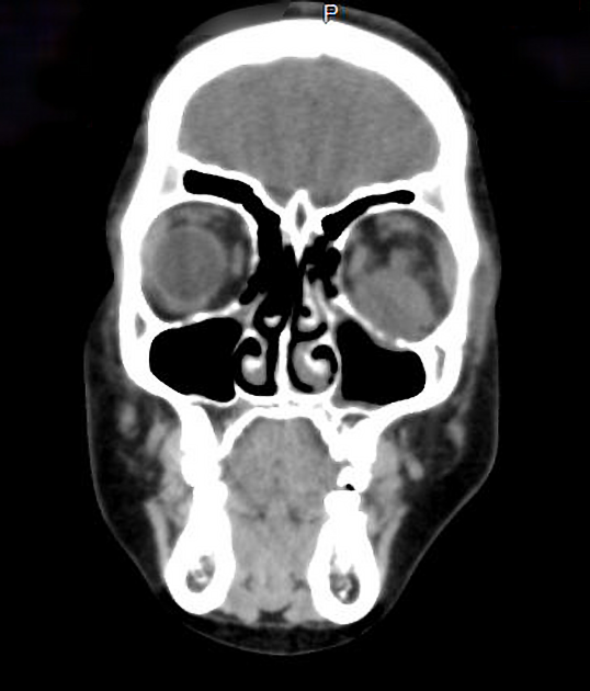
Clinical presentation is typically with a rapidly enlarging mass, often in the upper inner quadrant 1. It is usually painless but causes proptosis and diplopia. Often the mass invades the eyelid causing marked oedema 1.
Pathology
The vast majority of orbital rhabdomyosarcomas are of the embryonal subtype 1,3. Contrary to early belief, these tumours do not arise from the extraocular muscles, but rather develop from primitive mesenchymal cells that go on to differentiate into striated muscle cells 3.
Histologic subtypes:
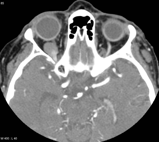
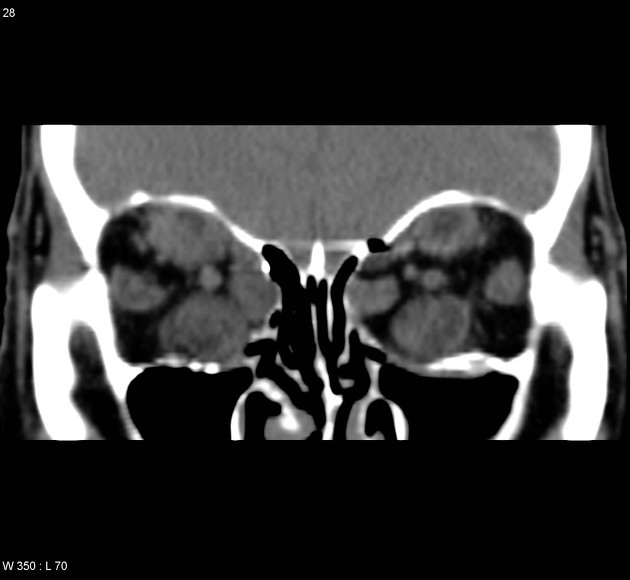
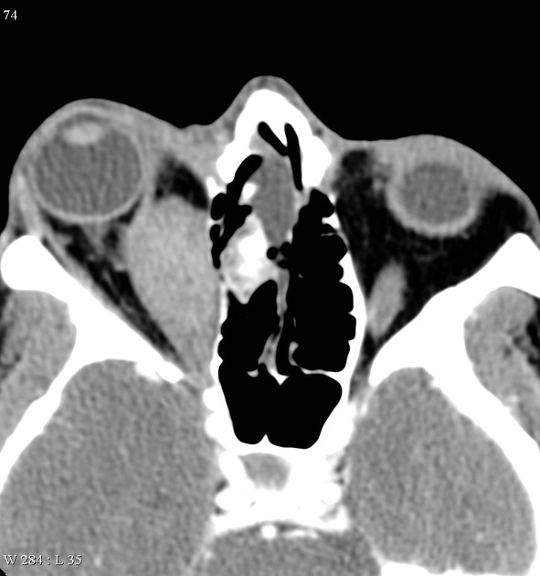
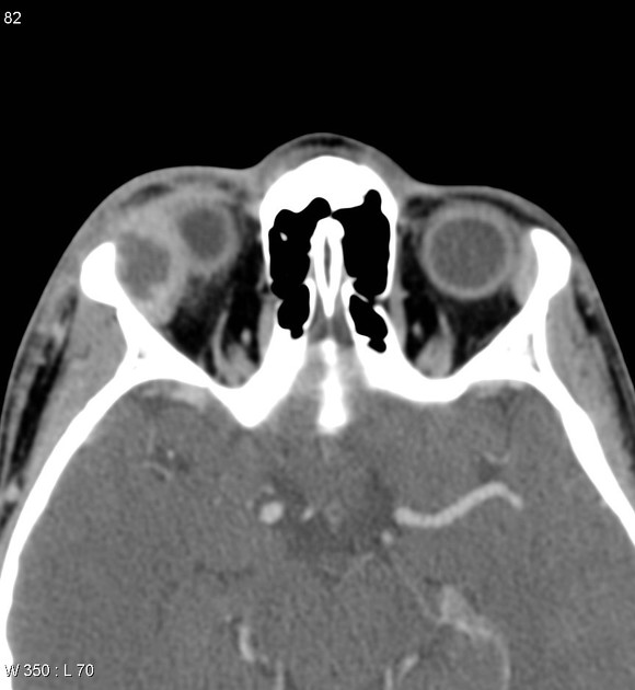
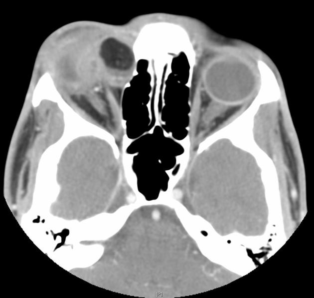
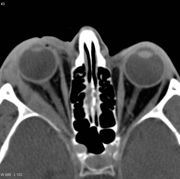
Radiographic features
CT and MRI are the modalities of choice for assessment of these masses, and to delineate adjacent structures.
It is important to report the location of the tumour epicentre as there is a correlation between location and histology: embryonal subtype more frequently arises in the superior orbit, whereas alveolar subtype is more common in the inferior orbit 3.
CT
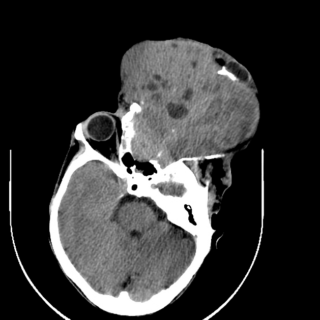
Rhabdomyosarcomas are typically homogeneous soft tissue masses isodense to normal muscle. The mass may extend into the eyelid or through the bone into the paranasal sinuses (especially the ethmoid sinus) and superiorly into the anterior cranial fossa.
Following contrast administration, enhancement is usually present.
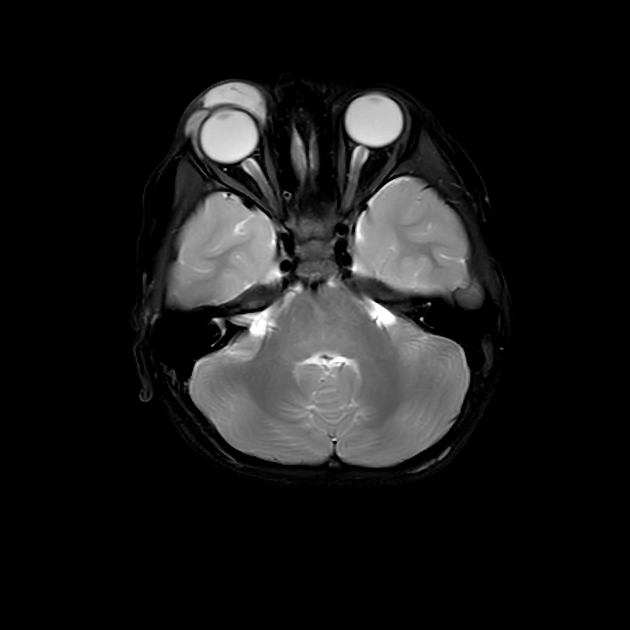
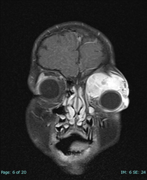
MRI
-
T1
low to intermediate intensity, isointense to adjacent muscle
areas of haemorrhage are common in alveolar and pleomorphic subtypes, but these are uncommon in the orbit 1
-
T2
usually hyperintense
T1 C+ (Gd): shows considerable enhancement
Treatment and prognosis
The mainstay of treatment is now a combination of radiotherapy and chemotherapy, which has achieved 5-year survival of over 90% for patients with embryonal rhabdomyosarcomas 2-3. In the small group of patients with alveolar rhabdomyosarcoma of the orbit, survival is lower but still good (75%) 3.
Radical surgery is no longer performed, with only a biopsy performed in many instances to confirm the diagnosis. (thus most tumours are Stage 1, Group III) 3. In some centres, extensive surgery is still performed to debulk the tumour 3.
Differential diagnosis
The differential is essentially that of an orbital mass, and includes 4-5:
-
tumours
schwannoma or neurofibroma
metastasis
orbital abscess
-
vascular lesions
capillary haemangioma: in infancy


 Unable to process the form. Check for errors and try again.