Citation, DOI, disclosures and article data
Citation:
Weerakkody Y, Campos A, Elfeky M, et al. Right ventricular dysfunction. Reference article, Radiopaedia.org (Accessed on 29 Mar 2025) https://doi.org/10.53347/rID-55495
Right ventricular dysfunction usually results from either pressure overload, volume overload, or a combination.
It occurs in a number of clinical scenarios, including:
It can manifest as right heart strain.
Sustained right ventricular dilatation and hypertrophy can frequently progress to right ventricular failure.
Right ventricular dysfunction can comprise of
Ultrasound
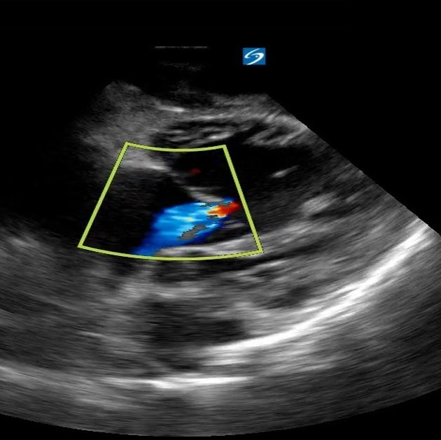
Echocardiography
Two dimensional echocardiography is usually considered the mainstay for analysis of right ventricular function.
Features include
right ventricular wall can be thickened (>4 mm) (often observed in congenital heart disease) or dilated (in acquired heart disease)
free wall may be hypokinetic; this is best appreciated on parasternal long axis projections
CT
May have a role in assessment. Suggestive signs include:
MRI
Several phenotypic patterns have been described 9.
pressure overload
volume overload
volume overload plus left ventricular dysfunction: considered 2nd commonest pattern
depressed biventricular function: commonest pattern
mixed overload, as there is co-existing biventricular dysfunction (in different degrees depending on disease duration), dilatation and right ventricular hypertrophy
-
1. Voelkel N, Quaife R, Leinwand L et al. Right Ventricular Function and Failure: Report of a National Heart, Lung, and Blood Institute Working Group on Cellular and Molecular Mechanisms of Right Heart Failure. Circulation. 2006;114(17):1883-91. doi:10.1161/CIRCULATIONAHA.106.632208 - Pubmed
-
2. Simon MA, Pinsky MR. Right ventricular dysfunction and failure in chronic pressure overload. Cardiology research and practice. 2011: 568095. doi:10.4061/2011/568095 - Pubmed
-
3. Bleeker GB, Steendijk P, Holman ER et-al. Acquired right ventricular dysfunction. Heart (British Cardiac Society). 92 Suppl 1: i14-8. doi:10.1136/hrt.2005.081547 - Pubmed
-
4. Hines R. Right Ventricular Function and Failure: A Review. Yale J Biol Med. 1991;64(4):295-307. PMC2589535 - Pubmed
-
5.Prognostic Value of Echocardiographic Right/Left Ventricular End-Diastolic Diameter Ratio in Patients With Acute Pulmonary Embolism
Frémont, Benoît et al.
CHEST , Volume 133 , Issue 2 , 358 - 362
-
6. Kang D, Thilo C, Schoepf U et al. CT Signs of Right Ventricular Dysfunction: Prognostic Role in Acute Pulmonary Embolism. JACC Cardiovasc Imaging. 2011;4(8):841-9. doi:10.1016/j.jcmg.2011.04.013 - Pubmed
-
7. Kang D, Ramos-Duran L, Schoepf U et al. Reproducibility of CT Signs of Right Ventricular Dysfunction in Acute Pulmonary Embolism. AJR Am J Roentgenol. 2010;194(6):1500-6. doi:10.2214/AJR.09.3717 - Pubmed
-
8. Boxt LM. Radiology of the right ventricle. Radiologic clinics of North America. 37 (2): 379-400. Pubmed
-
9. J Sánchez-Lázaro I, Almenar Bonet L, Igual Muñoz B et-al. Phenotypic patterns of right ventricular dysfunction: analysis by cardiac magnetic imaging. Heart international. 8 (1): e3. doi:10.4081/hi.2013.e3 - Pubmed
-
van der Meer R, Pattynama P, van Strijen M et al. Right Ventricular Dysfunction and Pulmonary Obstruction Index at Helical CT: Prediction of Clinical Outcome During 3-Month Follow-Up in Patients with Acute Pulmonary Embolism. Radiology. 2005;235(3):798-803. doi:10.1148/radiol.2353040593 - Pubmed
Promoted articles (advertising)


 Unable to process the form. Check for errors and try again.