
Spinal gangliogliomas are rare, comprising 1.1% of all spinal cord neoplasms 2. They are more frequent in children, representing 15% of intramedullary neoplasms in the pediatric age group 4.
This article specifically relates to spinal gangliogliomas. For a discussion on intracranial gangliogliomas and a general discussion of the pathology refer to the main article: ganglioglioma.
On this page:
Epidemiology
As is the case with intracranial variety, spinal gangliogliomas occur predominantly in children and young adults. The mean age of presentation is 19 years. There is no gender predilection.
Clinical presentation
Clinical presentation is similar to that of other intramedullary spinal tumors, with pain, weakness, and sensory changes common.
Pathology
Gangliogliomas are WHO grade I or II neoplasms 5.
Please refer to the main article: ganglioglioma.
Radiographic features
The most common location is the cervical cord, followed by the thoracic cord. Gangliogliomas may rarely involve the conus medullaris or the entire spinal cord (holocord presentation).
Plain radiograph/CT
As is the case with other intramedullary spinal tumors plain films and CT have little to offer in the diagnosis and characterization of spinal gangliogliomas. None the less a few features may be visible, including:
- scoliosis
- bony remodeling
- posterior vertebral body scalloping
- remodeling of the pedicle or posterior arch
- cord expansion may be visible on CT and vague enhancement may also be seen following contrast administration
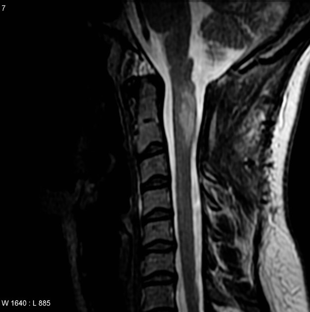
MRI
Gangliogliomas of the spinal cord typically involve long segments of the spinal cord, often extending for greater than eight vertebral body segments. They are commonly eccentric in location. Approximately half (46%) of these tumors contain tumoral cysts 3.
Signal characteristics include:
- T1: the mixed-signal intensity of gangliogliomas on T1 weighted images is due to the dual cellular elements of the tumor and is a unique finding among spinal cord tumors
-
T2
- high intensity
- surrounding edema is uncommon
- T1 C+ (Gd): most demonstrate patchy enhancement, however up to 15% cases may demonstrate no enhancement 2
- gradient echo: calcification is common, and will appear as areas of low signal with blooming
Treatment and prognosis
Gangliogliomas are generally slow-growing. Gross resection of the tumor is recommended. However, despite their low grade, there is a 27% rate of recurrence, which is approximately three to four times that of cerebral gangliogliomas, likely due to the increased difficulty of complete surgical excision of spinal tumors. Overall, there is an 89% 5-year survival rate after resection 3.
Differential diagnosis
The main differential diagnoses are spinal astrocytomas and spinal ependymomas. In many instances, a definite preoperative diagnosis is not possible as many of the features overlaps, however, some features are helpful in suggesting the diagnosis of a spinal ganglioglioma 6:
-
spinal astrocytoma
- difficult to distinguish from gangliogliomas
- majority enhance (used to be said that all enhance but this is not the case 7)
-
spinal ependymoma
- average length of four or less vertebral body segments (whereas gangliogliomas often extend over a greater distance than this)
- typically centrally located within the cord
- the vast majority enhance
- periapical cysts common
- hemosiderin capping common
-
transverse myelitis
- clinical presentation acuter
- absent bony remodeling
- central rather than eccentric
- rapid resolution on follow-up imaging


 Unable to process the form. Check for errors and try again.