
Superior ophthalmic vein thrombosis is rare but can potentially lead to visual loss in the affected eye(s).
On this page:
Epidemiology
Superior ophthalmic vein thrombosis is very rare, with an incidence of 3-4 cases/million/year 1. It can be either unilateral or bilateral.
Clinical presentation
Superior ophthalmic vein thrombosis may manifest as 1,2:
painful proptosis
conjunctival congestion
chemosis
visual disturbance which can progress to loss of vision
Complications are usually due to the underlying pathology 1.
Pathology
Etiologies can be divided into septic and aseptic 1,2:
-
septic etiologies
orbital cellulitis - most common cause
septic cavernous sinus thrombosis
-
aseptic etiologies
facial trauma
aseptic facial inflammation
hypercoagulable states
orbital neoplasm
Radiographic features
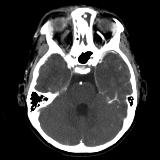
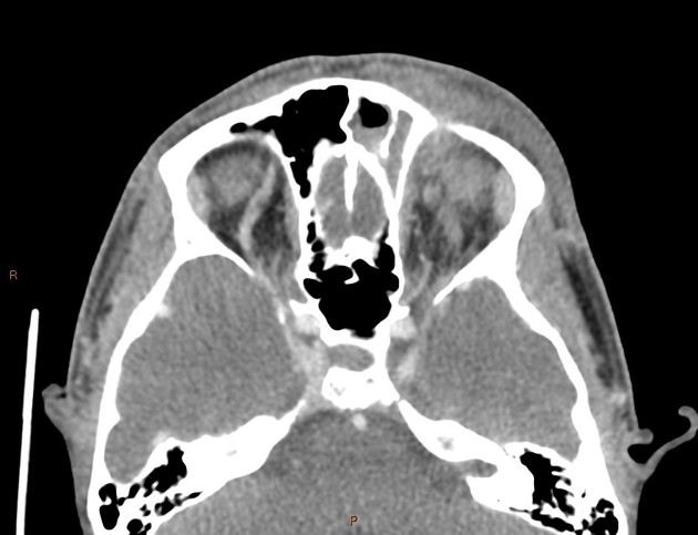
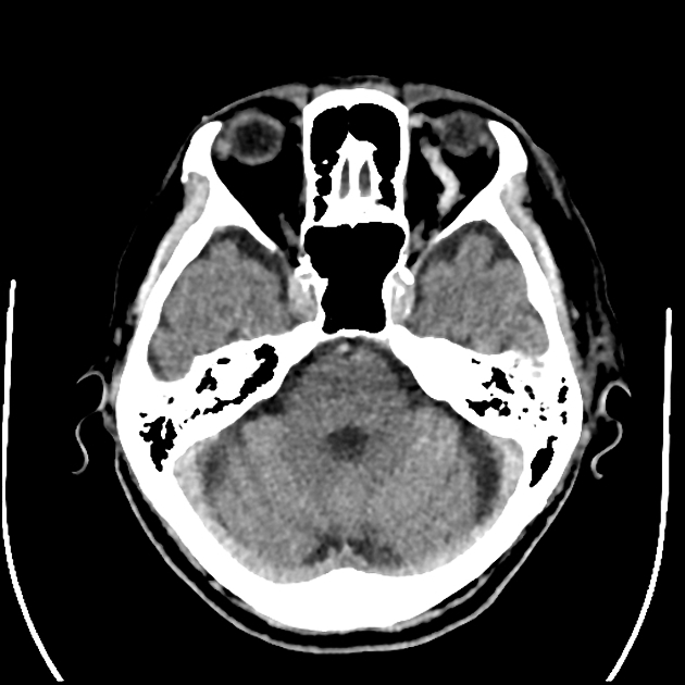
The modalities of choice for the diagnosis of superior ophthalmic vein thrombosis are CT venography (CTV) and MR venography (MRV). The thrombus is visualized as a linear filling defect that dilates the vein and can extend into the ipsilateral cavernous sinus (if its origin was not the cavernous sinus, to begin with).
Treatment and prognosis
Immediate anticoagulant treatment should be instituted, as well as treatment for the underlying cause where applicable.


 Unable to process the form. Check for errors and try again.