
Swirl sign (intracranial haemorrhage)
Citation, DOI, disclosures and article data
At the time the article was created Frank Gaillard had no recorded disclosures.
View Frank Gaillard's current disclosuresAt the time the article was last revised Kieran Kusel had no financial relationships to ineligible companies to disclose.
View Kieran Kusel's current disclosures- Swirl appearance in intracranial haemorrhage
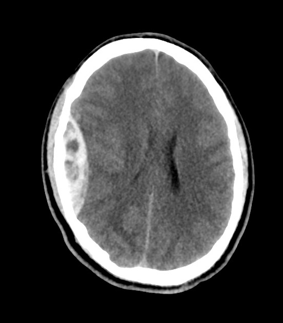
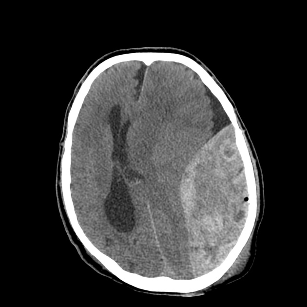
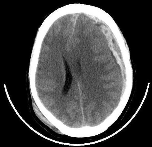
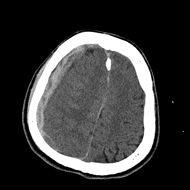
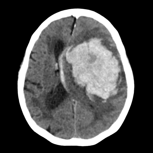
The swirl sign refers to the non-contrast CT appearance of acute extravasation of blood into a haematoma, for example an intracerebral haemorrhage, extradural haematoma or subdural haematoma. It represents unclotted fresh blood which is of lower attenuation than the clotted blood which surrounds it 1,5,6. It is in some ways the corollary of the spot sign on CTA which represents the same phenomenon, but in that case, caused by extraluminal focal accumulation/pooling/extravasation of contrast 2,5.
In an intracerebral haemorrhage, the swirl sign is one of many predictors of haematoma expansion 5 (see intracerebral haemorrhage for further discussion).
Terminology
The swirl sign is only one of many imaging features described in an attempt to predict haematoma growth, and in many instances, these signs overlap.
For example, the black hole sign is in many respects a swirl sign that is A) encapsulated by the haematoma and B) >28 HU lower in density than the surrounding haematoma 6.
Radiographic features
CT
The swirl sign denotes one or more areas within, or in continuity with, the haematoma that are of lower attenuation than the surrounding/adjacent clot. This can vary from isoattenuating to hypoattenuating to the adjacent brain 6.
Their morphology is variable; truly swirly (curvilinear, streaklike), rounded or branching 6.
Quiz questions

References
- 1. Al-Nakshabandi NA. The swirl sign. Radiology. 2001;218 (2): 433. doi:10.1148/radiology.218.2.r01fe09433 - Pubmed citation
- 2. Wada R, Aviv RI, Fox AJ et-al. CT angiography "spot sign" predicts hematoma expansion in acute intracerebral hemorrhage. Stroke. 2007;38 (4): 1257-62. doi:10.1161/01.STR.0000259633.59404.f3 - Pubmed citation
- 3. Das B, Khurana D, Ahuja CK. Bilateral "Swirl Sign": A predictor of rebleed. (2016) Annals of Indian Academy of Neurology. 19 (4): 514-515. doi:10.4103/0972-2327.194460 - Pubmed
- 4. Selariu E, Zia E, Brizzi M, Abul-Kasim K. Swirl sign in intracerebral haemorrhage: definition, prevalence, reliability and prognostic value. (2012) BMC neurology. 12: 109. doi:10.1186/1471-2377-12-109 - Pubmed
- 5. D. Ng, L. Churilov, P. Mitchell, R. Dowling, B. Yan. The CT Swirl Sign Is Associated with Hematoma Expansion in Intracerebral Hemorrhage. (2018) American Journal of Neuroradiology. doi:10.3174/ajnr.A5465 - Pubmed
- 6. Morotti A, Boulouis G, Dowlatshahi D et al. Standards for Detecting, Interpreting, and Reporting Noncontrast Computed Tomographic Markers of Intracerebral Hemorrhage Expansion. Ann Neurol. 2019;86(4):480-92. doi:10.1002/ana.25563 - Pubmed
Incoming Links
- Hyperacute epidural hematoma following ventriculoperitoneal shunt placement
- CT angiographic spot sign
- Acute epidural hematoma
- Bleeding butterfly glioma
- Rupture of anterior communicating artery aneurysm
- Coagulopathy related intracerebral hemorrhage on background of uremic encephalopathy
- Ruptured intracranial mycotic aneurysm
- Extradural hematoma
- Venous extradural haemorrhage at the vertex
- Sigmoid volvulus
- Lobar intracerebral haemorrhage
- Swirl sign of intracranial haemorrhage
- Extradural haematoma
- Extradural haematoma - swirl sign
- Subdural haematoma - swirl sign
- Extradural haematoma
Related articles: Stroke and intracranial haemorrhage
-
stroke and intracranial haemorrhage
- general articles
-
ischaemic stroke
- general discussions
- scoring and classification systems
- Alberta stroke program early CT score (ASPECTS)
- ASCOD classification
- Canadian Neurological Scale
- Heidelberg bleeding classification
- NIH Stroke Scale
- Mathew stroke scale
- modified Rankin scale
- Orgogozo Stroke Scale
- Scandinavian Stroke Scale
- thrombolysis in cerebral infarction (TICI) scale
- TOAST classification
- collateral vessel scores
- signs
- by region
- hemispheric infarcts
- frontal lobe infarct
- parietal lobe infarct
- temporal lobe infarct
- occipital lobe infarct
- alexia without agraphia syndrome: PCA
- cortical blindness syndrome (Anton syndrome): top of basilar or bilateral PCA
- Balint syndrome: bilateral PCA
- lacunar infarct
-
thalamic infarct
- artery of Percheron infarct
- Déjerine-Roussy syndrome (thalamic pain syndrome): thalamoperforators of PCA
- top of the basilar syndrome
- striatocapsular infarct
- choroid plexus infarct
- cerebellar infarct
-
brainstem infarct
- midbrain infarct
- Benedikt syndrome: PCA
- Claude syndrome: PCA
- Nothnagel syndrome: PCA
- Weber syndrome: PCA
- Wernekink commissure syndrome
- pontine infarct
- Brissaud-Sicard syndrome
- facial colliculus syndrome
- Gasperini syndrome: basilar artery or AICA
- inferior medial pontine syndrome (Foville syndrome): basilar artery
- lateral pontine syndrome (Marie-Foix syndrome): basilar artery or AICA
- locked-in syndrome: basilar artery
- Millard-Gubler syndrome: basilar artery
- Raymond syndrome: basilar artery
- medullary infarct
- Babinski-Nageotte syndrome
- Cestan-Chenais syndrome
- hemimedullary syndrome (Reinhold syndrome)
- lateral medullary stroke syndrome (Wallenberg syndrome)
- medial medullary syndrome (Déjerine syndrome)
- Opalski syndrome
- midbrain infarct
- acute spinal cord ischaemia syndrome
- hemispheric infarcts
- by vascular territory
- by vessel size
- treatment options
- complications
-
intracranial haemorrhage
-
intra-axial haemorrhage
- signs and formulas
- ABC/2 (volume estimation)
- black hole sign
- blend sign
- cashew nut sign
- CTA spot sign
- island sign
- satellite sign
- swirl sign
- zebra sign
- by type
- by location
- signs and formulas
- extra-axial haemorrhage
- extradural haemorrhage (EDH)
- intralaminar dural haemorrhage
- subdural haemorrhage (SDH)
-
subarachnoid haemorrhage (SAH)
- types
- complications
- grading systems
- subpial haemorrhage
-
intra-axial haemorrhage


 Unable to process the form. Check for errors and try again.