
Testicular appendages
Citation, DOI, disclosures and article data
At the time the article was created Praveen Jha had no recorded disclosures.
View Praveen Jha's current disclosuresAt the time the article was last revised Henry Knipe had the following disclosures:
- Micro-X Ltd, Shareholder (past)
These were assessed during peer review and were determined to not be relevant to the changes that were made.
View Henry Knipe's current disclosures- Testicular and epididymal appendages
Testicular and epididymal appendages are remnants of embryonic ducts and are quite common, with one or more being present in ~70% of patients 1.
Gross anatomy
Four such appendages have been described:
-
testicular appendix (hydatid of Morgagni)
- it is a Müllerian duct remnant (paramesonephric duct)
-
epididymal appendix
- it is a Wolffian duct remnant (mesonephric duct)
-
vas aberrans (of Haller)
- superior or inferior group of aberrant vessels located in a fissure between the epididymis and testis 1
-
paradidymis (Giraldes organ)
- vestigial structure located on the distal portion of the spermatic cord 1
Radiographic features
Ultrasound
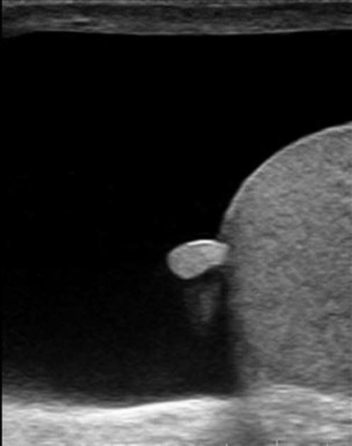
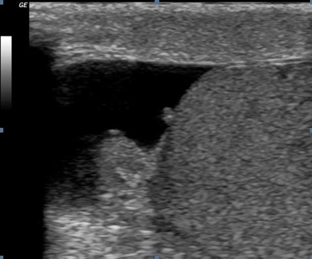
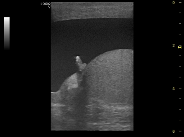
A testicular appendix may be seen between the testis and epididymis on normal sonography, in many instances. An epididymal appendix is attached to the head of the epididymis and may be seen during sonography in one or both testes. However, both vas aberrans and paradidymis are not usually seen during imaging.
References
- 1. Favorito L, Cavalcante A, Babinski M. Study on the Incidence of Testicular and Epididymal Appendages in Patients with Cryptorchidism. Int Braz J Urol. 2004;30(1):49-52. doi:10.1590/s1677-55382004000100011 - Pubmed
Incoming Links
Related articles: Anatomy: Abdominopelvic
- skeleton of the abdomen and pelvis
- muscles of the abdomen and pelvis
- spaces of the abdomen and pelvis
- anterior abdominal wall
- posterior abdominal wall
- abdominal cavity
- pelvic cavity
- perineum
- abdominal and pelvic viscera
- gastrointestinal tract
- spleen
- hepatobiliary system
-
endocrine system
-
adrenal gland
- adrenal vessels
- chromaffin cells
- variants
- pancreas
- organs of Zuckerkandl
-
adrenal gland
-
urinary system
-
kidney
- renal pelvis
- renal sinus
- avascular plane of Brodel
-
variants
- number
- fusion
- location
- shape
- ureter
- urinary bladder
- urethra
- embryology
-
kidney
- male reproductive system
-
female reproductive system
- vulva
- vagina
- uterus
- adnexa
- Fallopian tubes
- ovaries
- broad ligament (mnemonic)
- variant anatomy
- embryology
- blood supply of the abdomen and pelvis
- arteries
-
abdominal aorta
- inferior phrenic artery
- celiac artery
- superior mesenteric artery
- middle suprarenal artery
- renal artery (variant anatomy)
- gonadal artery (ovarian artery | testicular artery)
- inferior mesenteric artery
- lumbar arteries
- median sacral artery
-
common iliac artery
- external iliac artery
-
internal iliac artery (mnemonic)
- anterior division
- umbilical artery
- superior vesical artery
- obturator artery
- vaginal artery
- inferior vesical artery
- uterine artery
- middle rectal artery
-
internal pudendal artery
- inferior rectal artery
-
perineal artery
- posterior scrotal artery
- transverse perineal artery
- artery to the bulb
- deep artery of the penis/clitoris
- dorsal artery of the penis/clitoris
- inferior gluteal artery
- posterior division (mnemonic)
- variant anatomy
- anterior division
-
abdominal aorta
- portal venous system
- veins
- anastomoses
- arterioarterial anastomoses
- portal-systemic venous collateral pathways
- watershed areas
- arteries
- lymphatics
- innervation of the abdomen and pelvis
- thoracic splanchnic nerves
- lumbar plexus
-
sacral plexus
- lumbosacral trunk
- sciatic nerve
- superior gluteal nerve
- inferior gluteal nerve
- nerve to piriformis
- perforating cutaneous nerve
- posterior femoral cutaneous nerve
- parasympathetic pelvic splanchnic nerves
- pudendal nerve
- nerve to quadratus femoris and inferior gemellus muscles
- nerve to internal obturator and superior gemellus muscles
- autonomic ganglia and plexuses


 Unable to process the form. Check for errors and try again.