
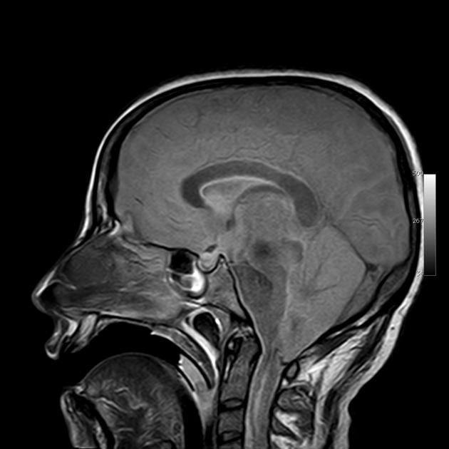
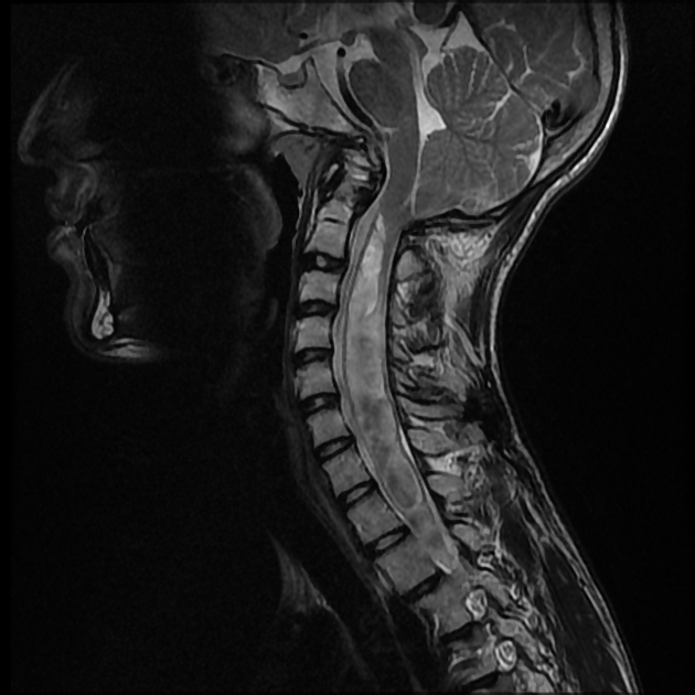
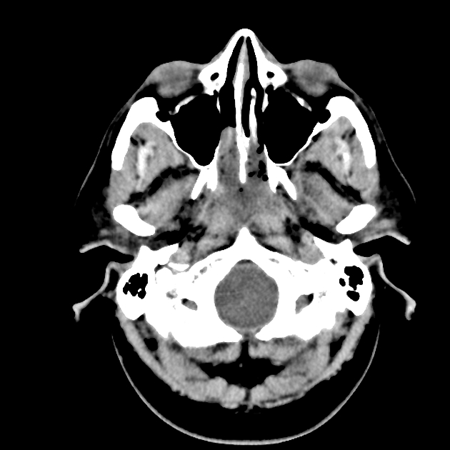
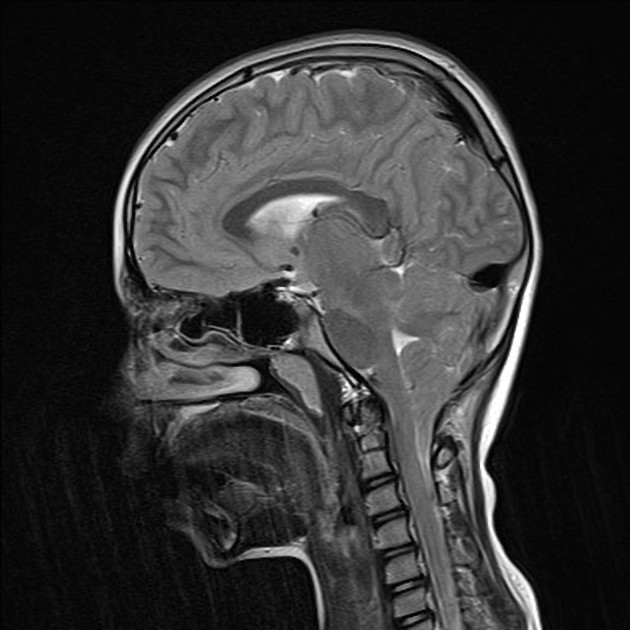
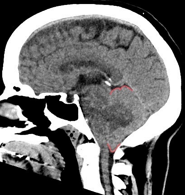
Tonsillar herniation is a type of brain herniation characterized by the inferior descent of the cerebellar tonsils below the foramen magnum 5. Clinically, the presence of tonsillar herniation is often called coning.
The terminology of caudally displaced tonsils is discussed in the article on cerebellar tonsillar ectopia.
Pathology
It is a secondary sign of markedly raised intracranial pressure. The skull is a rigid structure and an increase in volume of the contents is matched by a corresponding decrease in CSF volume, intravascular blood volume and/or brain herniation; see Monro-Kellie hypothesis. Common causes of raised intracranial pressure are tumor, hemorrhage, edema, stroke or abscess. Tonsillar descent is often a preterminal event as the brainstem is compressed against the clivus, affecting the vital life-sustaining functions of the pons and medulla, such as the respiratory and cardiac centers.
Radiographic features
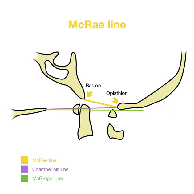
Tonsillar herniation is seen on CT and MRI as effacement of the CSF cisterns surrounding the brainstem and as inferior descent of the cerebellar tonsils >3 mm below foramen magnum level (McRae line)
Obstructive supratentorial hydrocephalus may result from fourth ventricle compression.


 Unable to process the form. Check for errors and try again.