Citation, DOI, disclosures and article data
Citation:
Feger J, Jones J, Roberts D, et al. Trochlear facet asymmetry. Reference article, Radiopaedia.org (Accessed on 25 Mar 2025) https://doi.org/10.53347/rID-78434
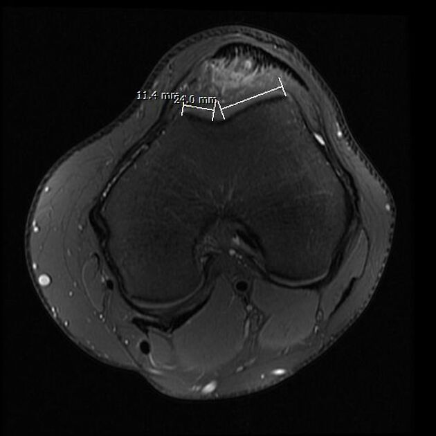
Trochlear facet asymmetry (FA) refers to the condition of the medial facet being abnormally small if compared to the lateral facet in trochlear dysplasia 1.
Facet asymmetry is also used as a measurement in magnet resonance imaging for trochlear dysplasia to make it more objective 1-3, this concept was adapted from axial radiographs 3.
One option to measure facet asymmetry is the ratio between medial versus lateral trochlear facet length calculated as (medial facet) / (lateral facet) 1. Another option is to calculate it the other way around, that is (lateral facet) / (medial facet) 2.
Facet asymmetry is traditionally measured 3 cm above the joint line 1,3.
A threshold of <0.4 or 40% was suggestive of trochlear dysplasia and showed a sensitivity and specificity of 100% and 96% in that study 2.
History and etymology
The principle has been adapted for magnetic resonance imaging by Pfirrmann 1.
ADVERTISEMENT: Supporters see fewer/no ads
-
1. Pfirrmann C, Zanetti M, Romero J, Hodler J. Femoral Trochlear Dysplasia: MR Findings. Radiology. 2000;216(3):858-64. doi:10.1148/radiology.216.3.r00se38858 - Pubmed
-
2. Charles M, Haloman S, Chen L, Ward S, Fithian D, Afra R. Magnetic Resonance Imaging–Based Topographical Differences Between Control and Recurrent Patellofemoral Instability Patients. Am J Sports Med. 2013;41(2):374-84. doi:10.1177/0363546512472441 - Pubmed
-
3. Ngai S, Smitaman E, Resnick D. Trochlear Dysplasia. Radsource – June 2018. MRI Web Clinic
-
4. Paiva M, Blønd L, Hölmich P et al. Quality Assessment of Radiological Measurements of Trochlear Dysplasia; a Literature Review. Knee Surg Sports Traumatol Arthrosc. 2017;26(3):746-55. doi:10.1007/s00167-017-4520-z - Pubmed
Promoted articles (advertising)


 Unable to process the form. Check for errors and try again.