
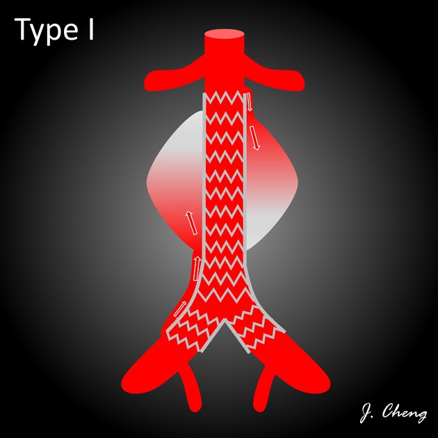
Type I endoleaks are a subgroup of endoleaks which occur at graft ends, often due to an inadequate seal.
On this page:
Epidemiology
These are considered a second most common type of endoleak with and estimated to account for 12% of all endoleaks according to one study 6.
Associations
most common after repair of thoracic aortic aneurysms 4
Pathology
They occur as a result of poor apposition between one of the attachment sites of a stent-graft and the native aortic or iliac artery wall. Blood can leak through this defect into the aneurysm sac.
They can be seen immediately after stent-graft deployment due to several reasons including
incomplete dilation of the stent-graft
aortic tortuosity
steep aortic angulation
Delayed type I endoleaks may be related to changes in the configuration of the aorta as the aneurysm sac shrinks. These are considered high-pressure endoleaks, and there is a high risk of aneurysm sac rupture because of direct exposure of the aneurysm wall to aortic pressure.
Subtypes
Type I endoleaks can be subdivided into three further categories:
Ia: proximal
Ib: distal
Ic: iliac occluder
Radiographic features
Type I endoleaks can be associated with measurable increases in aneurysm sac size.
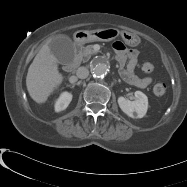
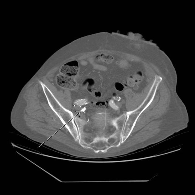
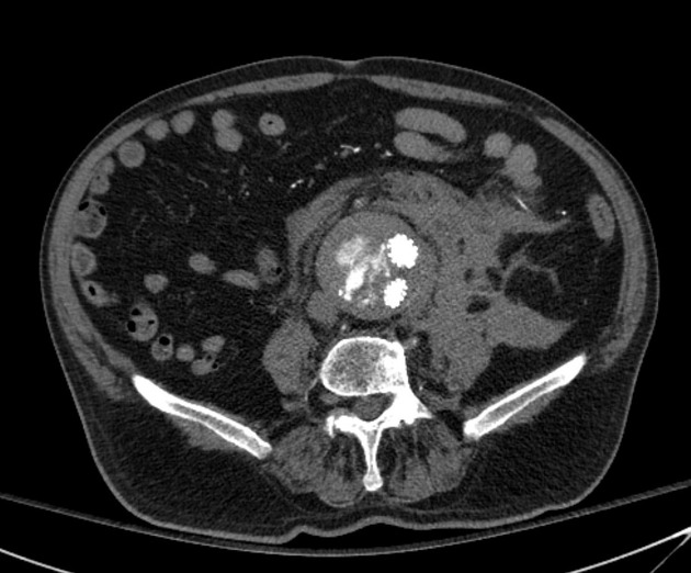
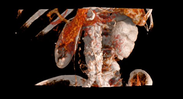
CT angiography
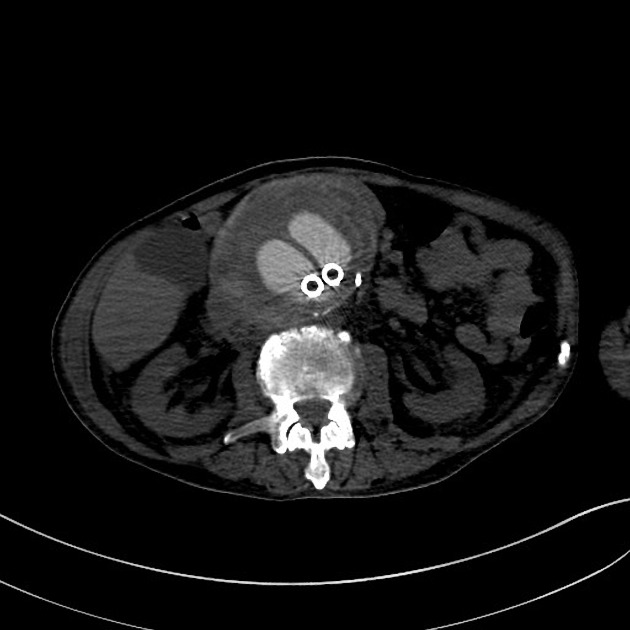
Preferred imaging modality. Multiphased scanning is helpful. Unenhanced CT may show hyperattenuating acute hemorrhage within the aneurysm sac.
After contrast administration, a dense contrast collection is usually seen centrally within the sac and is often continuous with one of the attachment sites.
Ultrasound
On Doppler sonography, a jet of flow may be seen originating from one of the attachment sites.
Treatment and prognosis
Type Ia leaks
Initial treatment can include balloon angioplasty of the proximal attachment site, aimed at remodeling the stent graft to achieve an adequate seal. If angioplasty is unsuccessful, balloon-expandable bare-metal stents such as Palmaz stents can be deployed over the affected attachment site to promote apposition of the proximal stent graft with the aortic wall.
In cases of undersized or poorly deployed endografts, covered extension cuffs can be used. These can be matched in size and material to the native endograft.
Some newer devices include EndoStaples and EndoAnchors which mechanically attach the proximal endograft with the aortic wall.
Late appearing type 1a endoleaks can have a higher rate of rupture 5.
Type Ib leaks
Are usually considered easier to manage than Ia leaks, with numerous available iliac extender limbs, covered stents, and bare-metal stents to close the endoleak defect.
Despite advances in endovascular techniques, there are still cases that require surgical repair for definitive treatment.


 Unable to process the form. Check for errors and try again.