Vascular imaging (dual-energy CT)
Citation, DOI & article data
- dual energy angiogram
Dual-energy CT has a variety of clinical applications in vascular imaging, due to the utilization of material decomposition and the ability to reconstruct images at a variety of energy levels.
Clinical applications
High kVp CT scans have lower contrast than that of lower kVp due to the K-edge of iodine, giving the lower energy of the dual energy scan an advantage over conventional CT. The attenuation values of large vessels enhanced with iodine are 70% higher at 80 kVp than at 140 kVp 1. In the event of suboptimal contrast injection and or timing, the lower energy set (closer to the K-edge of iodine) can be favored to improve the contrast resolution in various studies from pulmonary angiograms to aortograms 2.
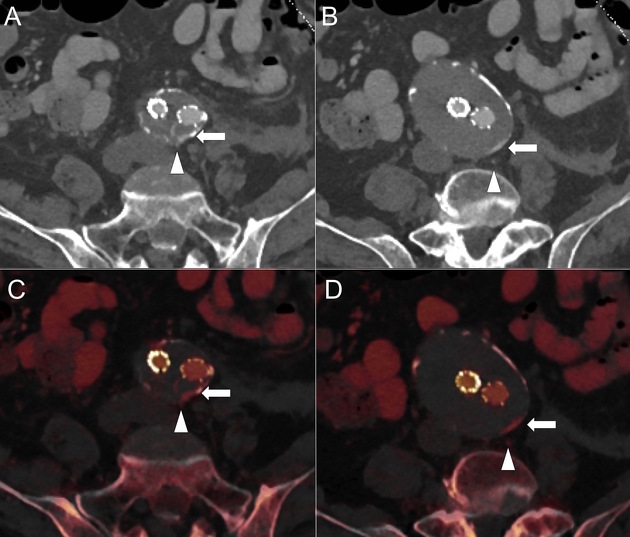
EVAR surveillance
Dual-energy aortogram in surveillance of endovascular aneurysm repair improves detection of endoleaks in fewer acquisitions 1; low kVp scanning can detect subtle leaks, while the virtual non-contrast images replace the unenhanced scan allowing a substantial reduction in radiation burden in patients that require life-long checkups 3,4.
Contrast sparing
Using lower energy data sets are proven to increase the arterial enhancement of pulmonary angiograms and other contrast studies due to the K-edge of iodine being closer to the lower energy used in a dual energy scanner 5-7.
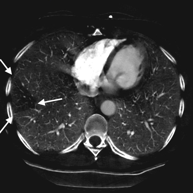
Pulmonary angiography
The 80 kVp image has the potential to improve subsegmental pulmonary artery perfusion and distal pulmonary embolus detection 2. In the setting of suboptimal enhancement of the pulmonary artery, virtual monochromatic reconstructions at lower energy levels can increase vessel enhancement, at the cost of increased noise.
Perfusion blood volume maps can be used to identify the segmental or subsegmental areas of lung affected by a pulmonary embolus. It is important to note that atelectasis, cardiac motion and streak artefact can all cause perfusion defects 10.
References
- 1. Vlahos I, Godoy MC, Naidich DP. Dual-energy computed tomography imaging of the aorta. J Thorac Imaging. 2010;25 (4): 289-300. doi:10.1097/RTI.0b013e3181dc2b4c - Pubmed citation
- 2. Godoy MC, Heller SL, Naidich DP et-al. Dual-energy MDCT: comparison of pulmonary artery enhancement on dedicated CT pulmonary angiography, routine and low contrast volume studies. Eur J Radiol. 2011;79 (2): e11-7. doi:10.1016/j.ejrad.2009.12.030 - Pubmed citation
- 3. Boll DT, Merkle EM, Paulson EK et-al. Coronary stent patency: dual-energy multidetector CT assessment in a pilot study with anthropomorphic phantom. Radiology. 2008;247 (3): 687-95. doi:10.1148/radiol.2473070849 - Pubmed citation
- 4. Aran S, Daftari Besheli L, Besheli LD et-al. Applications of dual-energy CT in emergency radiology. AJR Am J Roentgenol. 2014;202 (4): W314-24. doi:10.2214/AJR.13.11682 - Pubmed citation
- 5. Lu GM, Wu SY, Yeh BM et-al. Dual-energy computed tomography in pulmonary embolism. Br J Radiol. 2010;83 (992): 707-18. doi:10.1259/bjr/16337436 - Free text at pubmed - Pubmed citation
- 6. Ascenti G, Mazziotti S, Lamberto S, Bottari A, Caloggero S, Racchiusa S, Mileto A, Scribano E. Dual-energy CT for detection of endoleaks after endovascular abdominal aneurysm repair: usefulness of colored iodine overlay. Am J Roentgenol. 2011 Jun;196(6):1408-14. doi: 10.2214/AJR.10.4505.
- 7. Rutherford RA, Pullan BR, Isherwood I. Measurement of effective atomic number and electron density using an EMI scanner. Neuroradiology. 1976;11 (1): 15-21. Pubmed citation
- 8. Boll DT, Merkle EM, Paulson EK et-al. Coronary stent patency: dual-energy multidetector CT assessment in a pilot study with anthropomorphic phantom. Radiology. 2008;247 (3): 687-95. doi:10.1148/radiol.2473070849 - Pubmed citation
- 9. Murphy Andrew, Jeffrey Cheng and Jit Pratap et al. "Dual-Energy Computed Tomography Pulmonary Angiography: Comparison of Vessel Enhancement between Linear Blended and Virtual Monoenergetic Reconstruction Techniques". Journal of Medical Imaging and Radiation Sciences (2018). . doi:10.1016/j.jmir.2018.10.009
- 10. Lu GM, Wu SY, Yeh BM et-al. Dual-energy computed tomography in pulmonary embolism. Br J Radiol. 2010;83 (992): 707-18. doi:10.1259/bjr/16337436 - Free text at pubmed - Pubmed citation
Related articles: Imaging technology
- imaging technology
- imaging physics
- imaging in practice
-
x-rays
- x-ray physics
- x-ray in practice
- x-ray production
- x-ray tube
- filters
- automatic exposure control (AEC)
- beam collimators
- grids
- air gap technique
- cassette
- intensifying screen
- x-ray film
- image intensifier
- digital radiography
- digital image
- mammography
- x-ray artifacts
- radiation units
- radiation safety
- radiation detectors
- fluoroscopy
-
computed tomography (CT)
- CT physics
- CT in practice
- CT technology
- CT image reconstruction
- CT image quality
- CT dose
-
CT contrast media
-
iodinated contrast media
- agents
- water soluble
- water insoluble
- vicarious contrast material excretion
- iodinated contrast media adverse reactions
- agents
- non-iodinated contrast media
-
iodinated contrast media
-
CT artifacts
- patient-based artifacts
- physics-based artifacts
- hardware-based artifacts
- ring artifact
- tube arcing
- out of field artifact
- air bubble artifact
- helical and multichannel artifacts
- CT safety
- history of CT
-
MRI
- MRI physics
- MRI in practice
- MRI hardware
- signal processing
-
MRI pulse sequences (basics | abbreviations | parameters)
- T1 weighted image
- T2 weighted image
- proton density weighted image
- chemical exchange saturation transfer
- CSF flow studies
- diffusion weighted imaging (DWI)
- echo-planar pulse sequences
- fat-suppressed imaging sequences
- gradient echo sequences
- inversion recovery sequences
- metal artifact reduction sequence (MARS)
-
perfusion-weighted imaging
- techniques
- derived values
- saturation recovery sequences
- spin echo sequences
- spiral pulse sequences
- susceptibility-weighted imaging (SWI)
- T1 rho
- MR angiography (and venography)
-
MR spectroscopy (MRS)
- 2-hydroxyglutarate peak: resonates at 2.25 ppm
- alanine peak: resonates at 1.48 ppm
- choline peak: resonates at 3.2 ppm
- citrate peak: resonates at 2.6 ppm
- creatine peak: resonates at 3.0 ppm
- functional MRI (fMRI)
- gamma-aminobutyric acid (GABA) peak: resonates at 2.2-2.4 ppm
- glutamine-glutamate peak: resonates at 2.2-2.4 ppm
- Hunter's angle
- lactate peak: resonates at 1.3 ppm
- lipids peak: resonates at 1.3 ppm
- myoinositol peak: resonates at 3.5 ppm
- MR fingerprinting
- N-acetylaspartate (NAA) peak: resonates at 2.0 ppm
- propylene glycol peak: resonates at 1.13 ppm
-
MRI artifacts
- MRI hardware and room shielding
- MRI software
- patient and physiologic motion
- tissue heterogeneity and foreign bodies
- Fourier transform and Nyquist sampling theorem
- MRI contrast agents
- MRI safety
-
ultrasound
- ultrasound physics
-
transducers
- linear array
- convex array
- phased array
- frame averaging (frame persistence)
- ultrasound image resolution
- imaging modes and display
- pulse-echo imaging
- real-time imaging
-
Doppler imaging
- Doppler effect
- color Doppler
- power Doppler
- B flow
- color box
- Doppler angle
- pulse repetition frequency and scale
- wall filter
- color write priority
- packet size (dwell time)
- peak systolic velocity
- end-diastolic velocity
- resistive index
- pulsatility index
- Reynolds number
- panoramic imaging
- compound imaging
- harmonic imaging
- elastography
- scanning modes
- 2D ultrasound
- 3D ultrasound
- 4D ultrasound
- M-mode
-
ultrasound artifacts
- acoustic shadowing
- acoustic enhancement
- beam width artifact
- reverberation artifact
- ring down artifact
- mirror image artifact
- side lobe artifact
- speckle artifact
- speed displacement artifact
- refraction artifact
- multipath artifact
- anisotropy
- electrical interference artifact
- hardware-related artifacts
- Doppler artifacts
- aliasing
- tissue vibration
- spectral broadening
- blooming
- motion (flash) artifact
- twinkling artifact
- acoustic streaming
- biological effects of ultrasound
- history of ultrasound
-
nuclear medicine
- nuclear medicine physics
- detectors
- tissue to background ratio
-
radiopharmaceuticals
- fundamentals of radiopharmaceuticals
- radiopharmaceutical labeling
- radiopharmaceutical production
- nuclear reactor produced radionuclides
- cyclotron produced radionuclides
- radiation detection
- dosimetry
- specific agents
- carbon-11
- chromium-51
- fluorine agents
- gallium agents
- Ga-67 citrate
- Ga-68
- iodine agents
-
I-123
- I-123 iodide
- I-123 ioflupane (DaTSCAN)
- I-123 ortho-iodohippurate
- I-131
-
MIBG scans
- I-123 MIBG
- I-131 MIBG
-
I-123
- indium agents
- In-111 Octreoscan
- In-111 OncoScint
- In-111 Prostascint
- In-111 oxine labeled WBC
- krypton-81m
- nitrogen-13
- oxygen-15
- phosphorus-32
- selenium-75
-
technetium agents
- Tc-99m DMSA
- Tc-99m DTPA
- Tc-99m DTPA aerosol
- Tc-99m HMPAO
- Tc-99m HMPAO labeled WBC
- Tc-99m MAA
- Tc-99m MAG3
- Tc-99m MDP
- Tc-99m mercaptoacetyltriglycine
- Tc-99m pertechnetate
- Tc-99m labeled RBC
- Tc-99m sestamibi
- Tc-99m sulfur colloid
- Tc-99m sulfur colloid (oral)
- thallium-201 chloride
- xenon agents
- in vivo therapeutic agents
- pharmaceuticals used in nuclear medicine
-
emerging methods in medical imaging
- radiography
- phase-contrast imaging
- CT
- deep-learning reconstruction
- photon counting CT
- virtual non-contrast imaging
- ultrasound
- magnetomotive ultrasound (MMUS)
- superb microvascular imaging
- ultrafast Doppler imaging
- ultrasound localization microscopy
- MRI
- nuclear medicine
- total body PET system
- immuno-PET
- miscellaneous
- radiography