
Presentation
Pleuritic chest pain.
Patient Data
Age: 50 years
Gender: Female
From the case:
Dual-energy CTPA


Download
Info
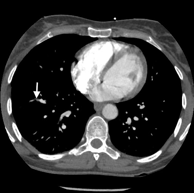
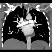
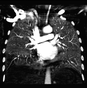
On conventional CTPA images, an apparent filling defect (arrow) is seen in a subsegmental right lower lobe pulmonary vessel. It was not immediately clear if this was in an artery or vein. The dual-energy scan allows acquisition of iodine perfusion maps at the same time as conventional CTPA, and it shows a wedge-shaped perfusion defect correlating to the position of the filling defect on CTPA. This increases the level of confidence that the abnormality seen represents a pulmonary embolism. The iodine perfusion defect very much resembles a ventilation/perfusion scan perfusion abnormality.


 Unable to process the form. Check for errors and try again.