
Presentation
Headache and blurring of vision.
Patient Data


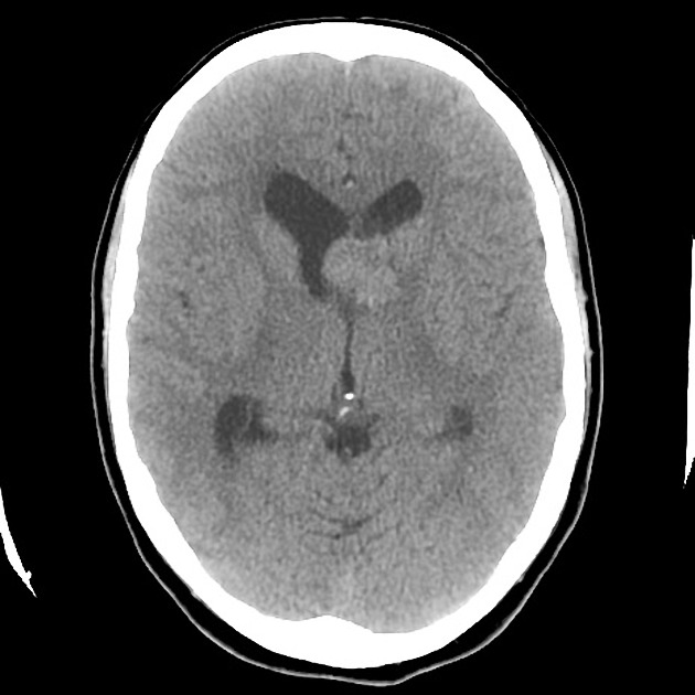
A 2 cm mass is located near the left foramen of Munro with associated ventriculomegaly, presumably due to obstruction at the level of the foramen. The mass is of soft tissue attenuation and demonstrates vivid contrast enhancement.
A single calcified subependymal focus is present on the lateral aspect of the right lateral ventricle. There is slight heterogeneity of the white matter of the cerebral hemispheres.
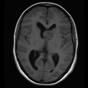
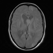
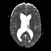
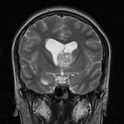

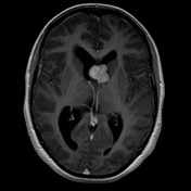
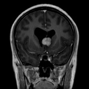
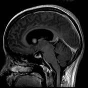

MRI demonstrates a vividly enhancing intraventricular mass in the left lateral ventricle resulting in a degree of obstructive non-communicating hydrocephalus (note the presence of ventriculomegaly and transependymal edema).
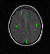
Widespread cortical and subcortical white matter regions of high T2 are seen as well as a number of prominent radial glial bands.
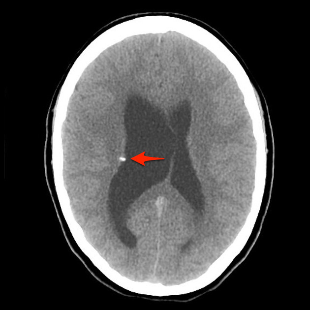
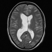
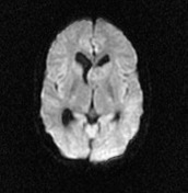
Calcified subependymal nodule (red arrow) is readily seen projecting into the lumen of the right lateral ventricle.

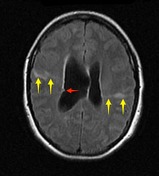
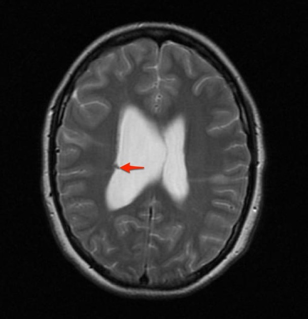
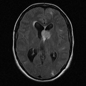
Subependymal nodule seen to be calcified on CT is visible (red arrow) on FLAIR and T2 as a region of low signal.
Best seen on FLAIR are radial glial bands (yellow arrows) and cortical / subcortical tubers (green arrows).
Case Discussion
This case demonstrates typical appearances of tuberous sclerosis with pathologicaly proven subependymal giant cell astrocytoma.


 Unable to process the form. Check for errors and try again.