
Presentation
Long-standing, vague abdominal pain for several months.
Patient Data


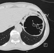
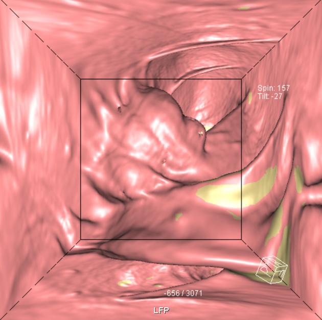
A multilobulated broad-based polypoid lesion was found at the splenic flexure. On the two-dimensional images the lesion demonstrates a rather ‘foamy’ appearance caused by multiple air-containing lobules.
Case Discussion
Patient presented with a history of vague abdominal pain for several months. Physical examination and laboratory tests were negative. Routine ultrasound examination showed no relevant abnormalities. The patient was not reassured and optical colonoscopy was performed to exclude colonic pathology. A polyp-like lesion with intact overlying mucosa was seen and interpreted as an external lesion; therefore no biopsy was taken. Consecutively a CT colonography was performed.
Pneumatosis cystoides coli (PCC) is a rare condition defined as an abnormal location of gas within the colonic wall and regarded as the colonic variant of pneumatosis cystoides intestinalis. It typically presents with multiple gas-filled cysts in the submucosa and/or subserosa of the colon. The size of the cysts may range from a few millimeters to several centimeters.
With CT it is possible to exclude other conditions such as pneumoperitoneum or gas in the portal venous system. CT colonography is the modality of choice in diagnosing PCC, allowing the exact location and configuration of the abnormalities to be described.
With acknowledgements to Dr. G. van der Horn


 Unable to process the form. Check for errors and try again.