
Note: This case has been tagged as "legacy" as it no longer meets image preparation and/or other case publication guidelines.
From the case:
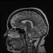
Suprasellar meningioma


Download
Info
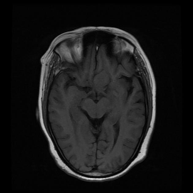
Isointense mass just left of suprasellar region with marked homogeneous enhancement.


 Unable to process the form. Check for errors and try again.