Presentation
Worsening gait and cerebellar signs
Patient Data
Age: 6 years
Note: This case has been tagged as "legacy" as it no longer meets image preparation and/or other case publication guidelines.
From the case:
Wilson disease
![](https://prod-images-static.radiopaedia.org/images/584070/560f192bf46d1b3e3c66d0cebe58de_thumb.jpg)
![](https://prod-images-static.radiopaedia.org/images/584076/c1ca4e0f791657f2293e5b16ec4494_thumb.jpg)
![](https://prod-images-static.radiopaedia.org/images/584088/93fbf0e17dbec2c58e921e406e2e36_thumb.jpg)
![](https://prod-images-static.radiopaedia.org/images/584082/86657397e026efba49f3ca5a03ee8e_thumb.jpg)
![](https://prod-images-static.radiopaedia.org/images/584094/14e1449c646f72a3eb784ef46abd58_thumb.jpg)
![](https://prod-images-static.radiopaedia.org/images/584106/ad99f01022a4bf590b745f1630f668_thumb.jpg)
![](https://prod-images-static.radiopaedia.org/images/584100/defd43e6159d6c84a22c690ddf2d7d_thumb.jpg)
![](https://prod-images-static.radiopaedia.org/images/584112/c8d661433049177c6df9f92029e5ce_thumb.jpg)
![](https://prod-images-static.radiopaedia.org/images/584118/76adae2ef53b164b0da392875ded69_thumb.jpg)
![](https://prod-images-static.radiopaedia.org/images/584124/0377075e89e0aac2da290b25f22978_thumb.jpg)
Download
Info
![](https://prod-images-static.radiopaedia.org/images/584070/560f192bf46d1b3e3c66d0cebe58de_big_gallery.jpg)
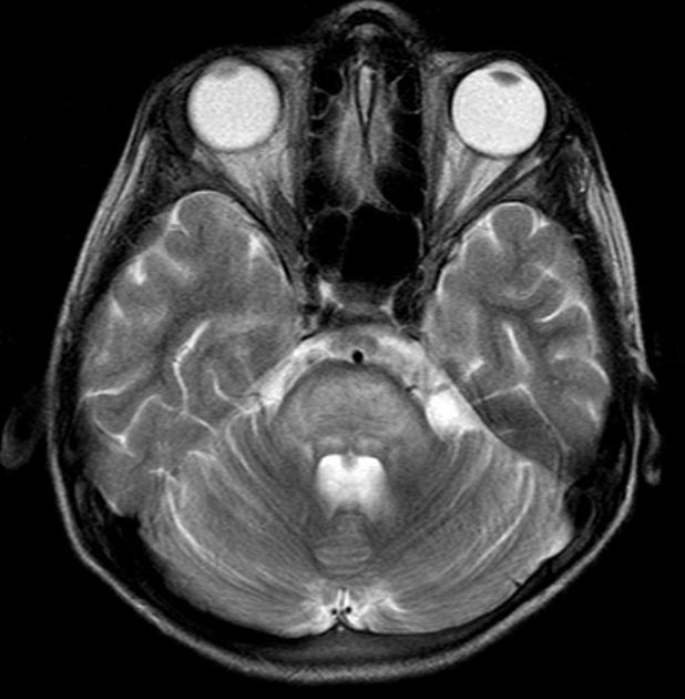
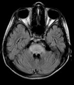
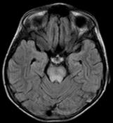
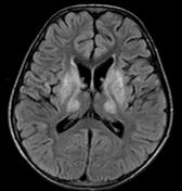
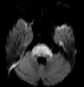
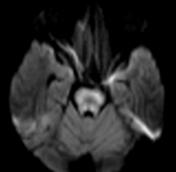
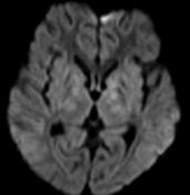
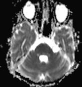
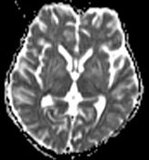
Diffuse T2 hyperintensities and restricted DWI signal of the pons with extension into the middle cerebellar peduncles. Also similar increased T2 signal of the symmetric basal ganglia, including the caudate nuclei, putamina, as well as the ventrolateral thalami, without the restriction of diffusion.
Case Discussion
Typical CNS appearances of Wilson disease, which was proven.