
Presentation
Infective symptoms.
Patient Data
Age: 75 years
Gender: Female
Download
Info
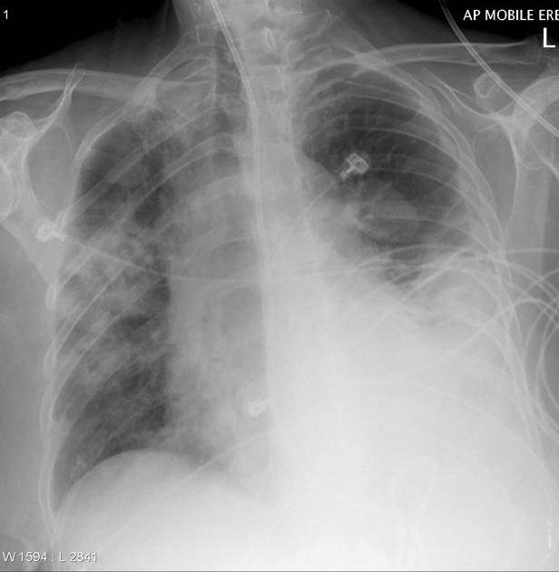
Chest x-ray (rotated to the right) demonstrates widespread airspace opacity (possibly with early cavitation) in the right mid zone. A large left sided pleural effusion is present with associated atelectasis. Nasogastric tube and ECG leads noted.
In the left upper zone a well circumscribed opacity is visible.
Download
Info
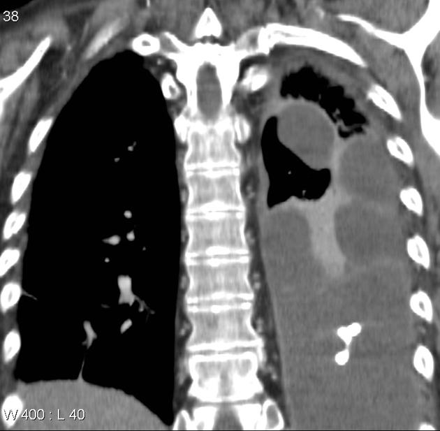
Coronal reformats of a CT of the chest obtained a few days earlier demonstrates that the opacity in the left upper zone is due to encysted fluid in the superior part of the oblique fissure.
Case Discussion
This case illustrates the typical appearance of encysted pleural fluid, resulting in a pulmonary pseudotumor. It also highlights the usefulness and necessity of reviewing previous films.


 Unable to process the form. Check for errors and try again.