
Presentation
Pain and difficulty urinating.
Patient Data
Age: 40 years
Gender: Female
From the case:
Adenocarcinoma arising in a urethral diverticulum


Download
Info
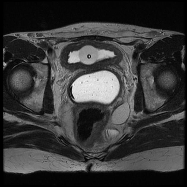
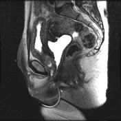
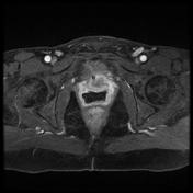
Vaginal contrast was used for this study.
Axial and coronal MR images of the pelvis demonstrate a circumferential mass around the urethra with extension into the posterolateral anterior vaginal wall. Additionally, superior extension into the bladder base is best seen on the coronal images.
Also, note the abnormal T2 signal above and below the mass in the periurethral regions suggesting a urethral diverticulum.
Case Discussion
This case is an opportunity to review periurethral mass lesions:
Differential considerations
- carcinoma arising from urethral diverticulum
- vaginal carcinoma
- vaginal metastasis
Diagnosis
Adenocarcinoma arising from a urethral diverticulum


 Unable to process the form. Check for errors and try again.