
Presentation
Adult presenting with knee pain


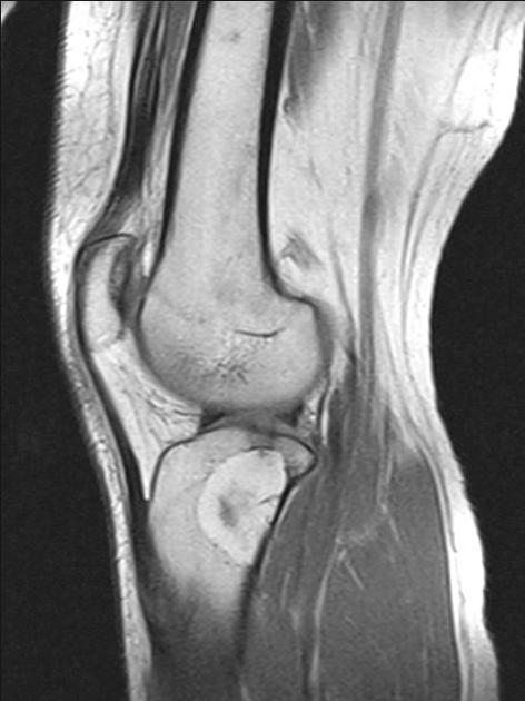
A well-delineated intraosseous lesion in the proximal tibia, which returns signal similar to fat on T1 and STIR. A focal central high signal on STIR suggests the presence of fluid raising suspicion of cystic degeneration component.
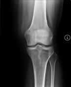
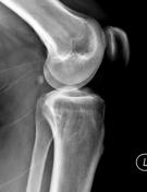

Well delineated lucent lesion within the proximal tibia.
Case Discussion
Intraosseous lipomas can contain varying amounts of fat, fibrous tissue, and cystic degeneration. This results in a range of radiographic manifestations. Stage 2 lesions demonstrate fat and fat necrosis, and also dystrophic calcification.
The characteristic fatty component that is isointense with fatty tissues on T1 images, occupies the periphery of the lesion. Central region which hyperintense on STIR is suggestive of fluid, which may represent cystic degeneration.


 Unable to process the form. Check for errors and try again.