
Presentation
Sudden onset abdominal pain, hypotension and tachycardia.
Patient Data


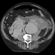
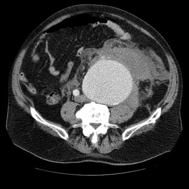
Contrast-enhanced 4-slice CT scan of the whole aorta (only the abdominal slices are shown) in the arterial phase and subsequent scan of the abdomen 2 min. post administration of i.v. contrast.
The main finding of the arterial phase is a ruptured aneurysm of the left common iliac artery which extends into the external iliac artery. It has a maximum diameter of 10 cm and extends over a length of 14 cm. Active extravasation of contrast-enhanced blood can be seen into the large retroperitoneal hematoma.
The subsequent scan reveals massive continuing bleeding and with a notable increase of the size of the retroperitoneal hematoma which now also involves the contralateral retroperitoneum and measures about 20 x 10 x 26 cm.
Case Discussion
This case demonstrates typical appearances of massive retroperitoneal hemorrhage from an uncommon cause: an isolated common iliac artery aneurysm - most patients would also have an abdominal aortic aneurysm.
The patient underwent emergent surgical intervention with the successful replacement of the iliac artery and eventual recovery. Most patients with ruptured aneurysms would not be so lucky.


 Unable to process the form. Check for errors and try again.