
Presentation
Severe headache and loss of vision of the left eye
Patient Data
Note: This case has been tagged as "legacy" as it no longer meets image preparation and/or other case publication guidelines.


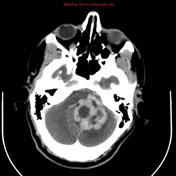
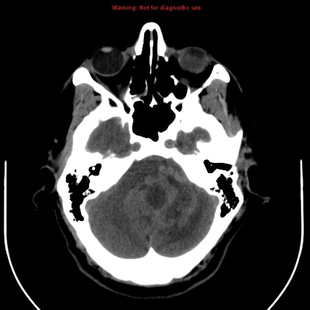
A large left cerebellar irregular mass lesion with necrotic core and irregular thick enhancing margins with prominent feeding arteries and draining veins. It is compressing and involving the left side of the 4th ventricle. Moderate supratentorial hydrocephalus is noted. It is involving the left cerebellar tonsil extending downwards compressing the cervicomedullary junction and brainstem.

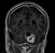
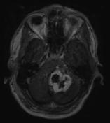
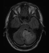
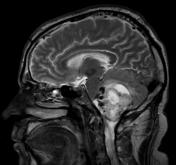
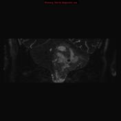
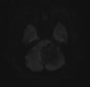
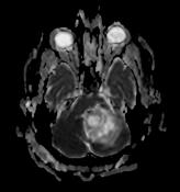

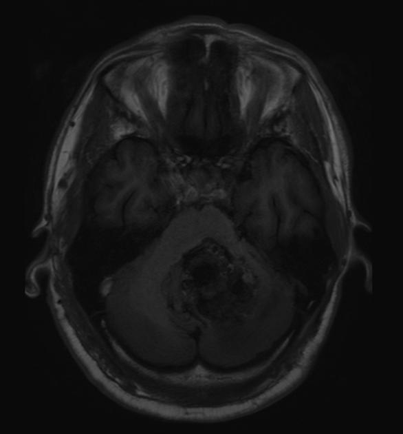
Left cerebellar lesion with low T1 signal, high T2 heterogeneous signal with avid post-contrast enhancement and necrotic core. Thin T2 cuts show multiple flow void channels inside and around the lesion.
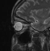
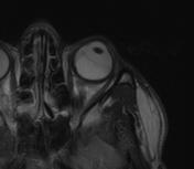
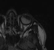
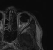
Left eye shows high T2 and FLAIR signal of the posterior part of vitreous body with low T1 signal, in keeping with hyperacute vitreous hemorrhage.
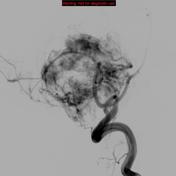
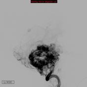
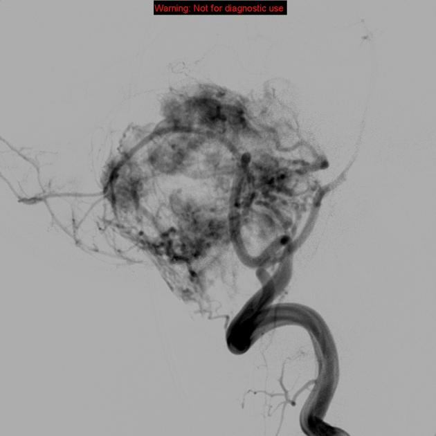
Left vertebral angiogram shows highly vascular lesion with prominent feeding arteries and draining veins.
Case Discussion
Pathologically proved hemangioblastoma. The differential diagnosis is astrocytoma.


 Unable to process the form. Check for errors and try again.