
Presentation
Left breast mass.
Patient Data
Age: 50 years
Gender: Female
From the case:
Breast carcinoma - BIRADS V


Download
Info
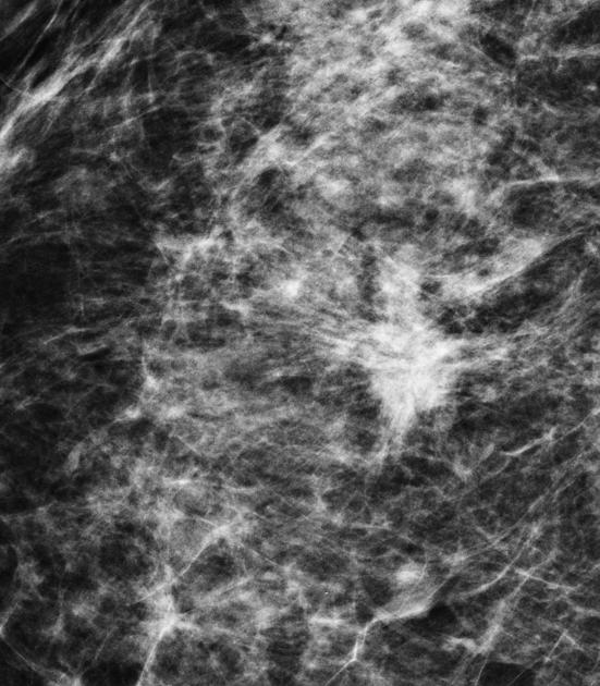
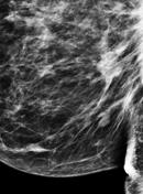
An irregular, dense, spiculated mass lesion. The lesion became more conspicuous on post compression view and it is associated with architectural distortion. No suspicious microcalcification is noted.
From the case:
Breast carcinoma - BIRADS V
Download
Info

Hypoechoic lesion, with irregular and angular margins, taller than wider, having thick echogenic capsule and demonstrates posterior shadowing. Vascularity is noted on color Doppler scan.
Case Discussion
The above mentioned findings are in keeping with typical BIRAD V lesion.


 Unable to process the form. Check for errors and try again.