
Presentation
Lower abdominal pain and fever.
Patient Data
Age: 30 years
Gender: Female
From the case:
Appendiceal abscess


Download
Info
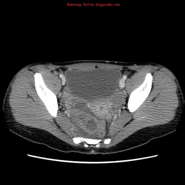
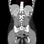
CT of the abdomen demonstrates a markedly dilated and inflamed, fluid filled tubular structure in the right lower pelvis which displaces the uterus and rectum.
CT imaging differential includes an appendiceal abscess and a tubo-ovarian abscess.
From the case:
Appendiceal abscess
Download
Info

Ultrasound demonstrates that this is separate from the normal looking right ovary and confirms the relationship with the cecum. Diameter: 3.8cm.
Case Discussion
Final diagnosis: Appendiceal abscess.


 Unable to process the form. Check for errors and try again.