
Presentation
Painless left infraclavicular chest wall mass for a few years.
Patient Data
Age: 50 years
Gender: Male
From the case:
Chest wall lipoma


Download
Info
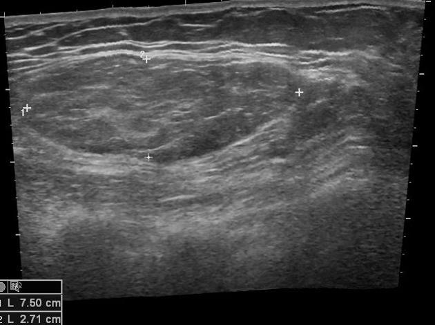
There is a well-defined, encapsulated mass with echopattern identical to fat at the site of swelling pointed by the patient. It is in the anterior chest wall muscle, deep to the subcutaneous fat. It is hyperechoic to overlying subcutaneous fat with the presence of echogenic incomplete lines. The lesion shows mild compressibility. There is no calcification/ cystic change/ vascularity in the lesion.
Case Discussion
The case shows a typical ultrasound appearance of the soft tissue lipoma in the anterior chest wall.


 Unable to process the form. Check for errors and try again.