
Presentation
Presenting with bleeding in 3rd trimester of pregnancy.
Patient Data
Age: 30 years
Gender: Female
From the case:
Placenta previa with vasa previa


Download
Info

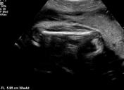
Selected images demonstrate that the placental leading edge reaches the internal cervical os (placenta previa). The umbilical vessels leave off from the placental leading edge and cross the internal os prior to entering the amniotic cavity (vasa previa with either marginal or velamentous cord insertion).
Selected images demonstrating normal biometry.
Case Discussion
Ultrasound demonstrates a placenta previa and vasa previa due to either marginal or velamentous cord insertion.


 Unable to process the form. Check for errors and try again.