
Presentation
Previously well. Sudden headache, vomiting and left sided weakness.
Patient Data
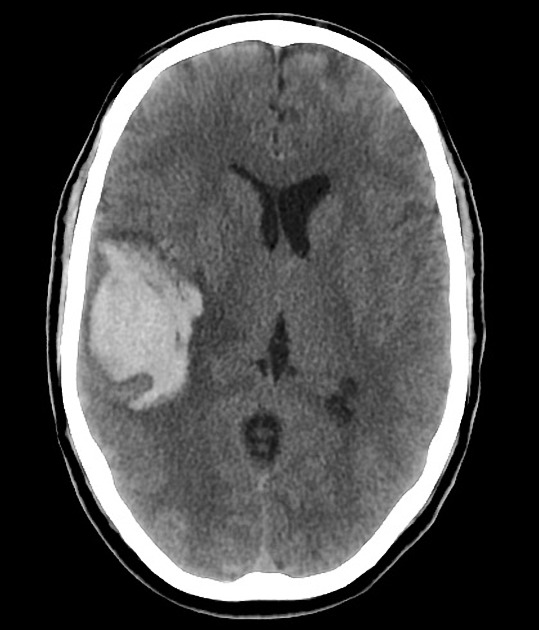
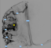
Non-contrast CT of the brain demonstrates a large right temporo-parietal hemorrhage. It is associated with moderate midline shift.
Anterior and posterior to the hematoma are nodular regions of lower attenuation (lower than the thrombus, similar to dural venous sinuses) with a single calcification.
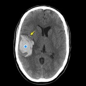
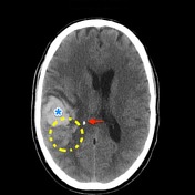
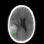
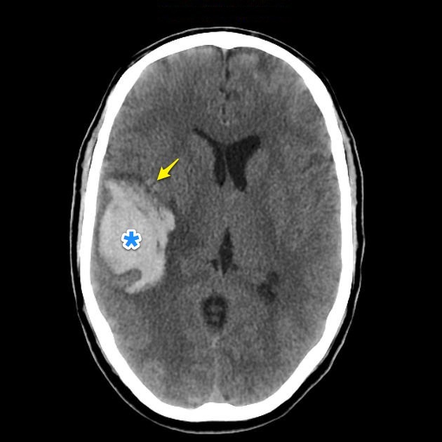
Large right temporo-parietal hemorrhage (*). Anterior and posterior to the hematoma are nodular regions of lower attenuation (yellow - lower attenuation than the thrombus, similar to dural venous sinuses) with a single calcification (green arrow) which is separate form the displaced choroid plexus calcification (red arrow).


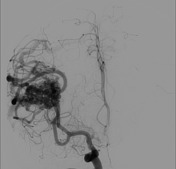
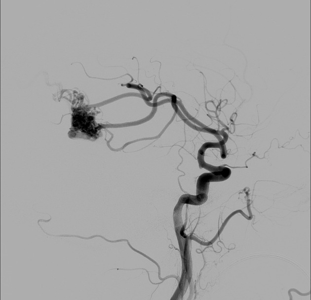
Right common carotid artery angiogram demonstrates a ~1.5cm nidus supplied predominantly by the right middle cerebral artery. The AVM has superficial drainage via markedly enlarged cortical veins, which drain in the dural sinuses. No deep drainage was identified.
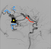
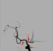
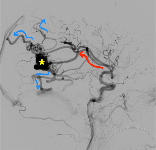
The nidus (*) is supplied by middle cerebral artery (MCA) branches (red) and drains via numerous enlarged cortical veins (blue CV).
Case Discussion
This case illustrates the typical and relatively subtle appearances of a cerebral AVM on non-contrast CT in the setting of a hemorrhage. An underlying lesion should always be suspected and sought when a young patient presents with a hemorrhage.


 Unable to process the form. Check for errors and try again.