
Presentation
Pain and deformity of the wrist.
Patient Data


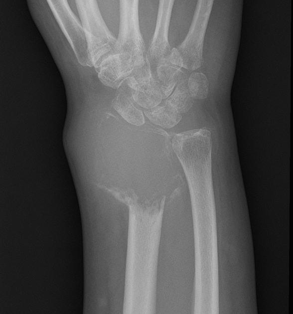
X-rays of the right forearm and wrist demonstrate a lytic expansile lesion involving the distal epiphysis of the radius. It abuts the radiocarpal joint (involving the epiphysis) and results in dorsal subluxation of the distal ulna. There is no matrix calcification. The margins are irregular with an aggressive appearance and relatively broad zone of transition. There is no convincing periosteal reaction. The carpus and ulnar appear unremarkable other than osteopaenic, presumably due to disuse.


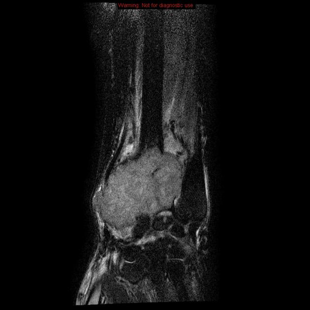
MRI confirms the plain film features. A mass is centered on the distal radius, involving the articular surface of the radiocarpal joint. The mass is solid with intermediate to high T2 signal and low T1 signal and demonstrates relatively homogeneous contrast enhancement. No cystic areas or fluid-fluid levels identified.
High T2 signal (edema) is seen extending proximally along the interosseous membrane with some fluid also seen within the carpus, but no significant bone marrow edema. The bone marrow of the more proximal radius, the ulnar and the carpal bones are unremarkable.
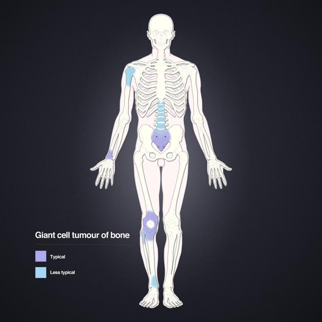
Distribution of giant cell tumors (GCT) of bone. Layout and distribution: Frank Gaillard 2009, Line drawing of skeleton: Patrick Lynch 2006, Creative Common NC-SA-BY
Case Discussion
This case illustrates typical appearances of a giant cell tumor (GCT).


 Unable to process the form. Check for errors and try again.