
Presentation
No history provided.
Patient Data
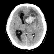
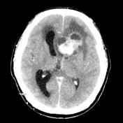

Digitized images from printed film. A large, heterogeneous mass is present within the left frontal lobe. It is strikingly hyperdense, without definite calcification. The solid component enhances. Hydrocephalus is present, best seen on the right, due to outflow obstruction.


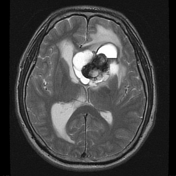
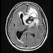
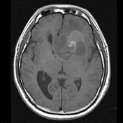
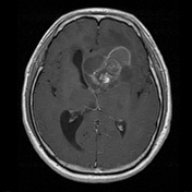
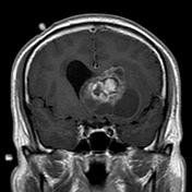
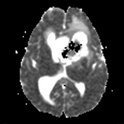
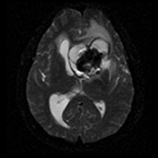
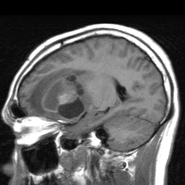
There is a large left frontal mixed cystic/solid lesion, measuring approximately 4x4x5cm in perpendicular dimensions, with intralesional hemorrhage and fluid level which may indicate areas of necrosis. Large surrounding vasogenic edema involving the contralateral hemisphere, and significant mass effect causing almost 2cm midline shift, obstructing the monro foramina resulting moderate non-communicating hydrocephalus.
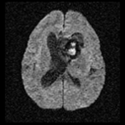
The epicenter of the mass demonstrates diffusion restriction. There is no evidence of an increased rCBV. MRS demonstrates significantly increased choline and lactate/lipid peak with substantial decrease in NAA (not shown).
The patient went on to have a craniotomy and excision of this lesion.

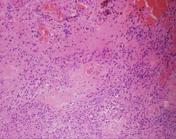
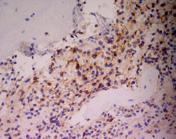
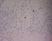
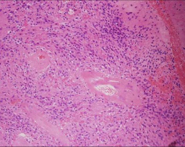
Paraffin sections show a moderately hypercellular glial tumor. Tumor cells have uniform round and ovoid nuclei with a granular arrangement of chromatin and a variable amount of pale to clear cytoplasm. These are arranged in prominent perivascular pseudorosettes as well as diffuse sheets. No mitotic figures are identified. A single small focus of vascular endothelial cell hyperpklasia is noted in one fragment. There is no necrosis. Large caliber intensely congested vascular channels are present in separate fragments. The majority of tumor cells show prominent perinuclear dot immunostaining for epithelial membrane antigen (EMA) typical of ependymal cells. There is also patchy immunostaining for GFAP. The features are of ependymoma. The topoisomerase labeling index is <1%.
DIAGNOSIS: Left frontal and lateral ventricle tumor: Ependymoma (WHO Grade II).
Case Discussion
This case highlights the difficulty in establishing a firm imaging diagnosis in atypical cases.


 Unable to process the form. Check for errors and try again.