
Presentation
Smoker. Weight loss.
Patient Data
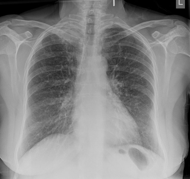
5cm right apical mass. No rib destruction.
Heart size normal. Lungs clear.
Normal mediastinal contours.


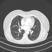
5cm spiculated mass in the apical segment of the right upper lobe with tethering to the pleural surface. No satellite nodules.
No supraclavicular or mediastinal lymphadenopathy.
No metastatic disease in the abdomen or pelvis.
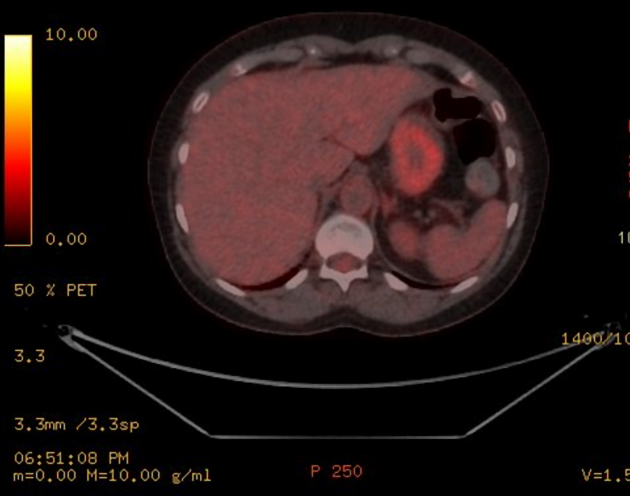
5cm FDG avid right upper lobe apical segment mass.
No locoregional or distant metastatic disease.
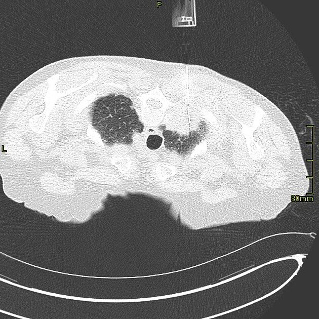
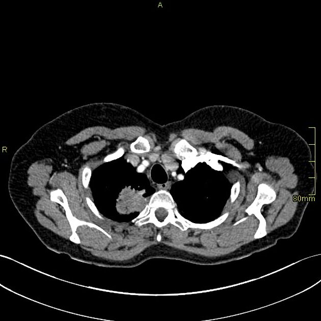
Prone position.
18G core biopsy of right apical mass.
Case Discussion
This patient encounter illustrates the steps in the diagnosis, work up and histological confirmation with image guided biopsy of a patient with potentially surgically curative lung cancer.
It also illustrates a classical example of a Pancoast tumor.
This was a confirmed lung malignancy that proceeded to surgery.


 Unable to process the form. Check for errors and try again.