Presentation
Blunt trauma to the left upper chest 3 days prior to the presentation. Painful shoulder and neck movements.
Patient Data
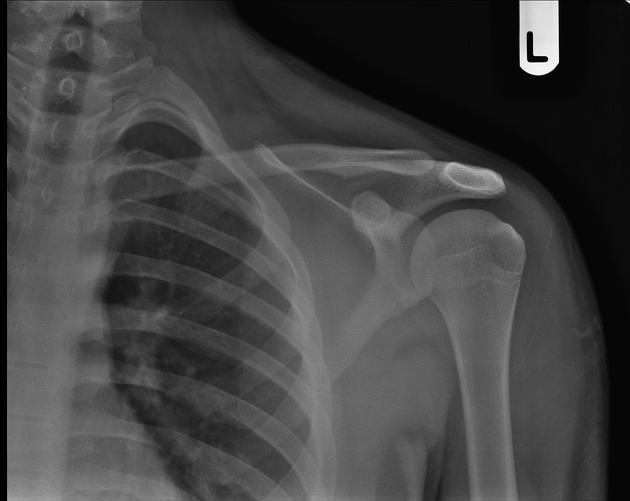
No fracture. Normal glenohumeral joint and acromioclavicular joint.

Posterior dislocation of the medial end of the left clavicle. Axial images reveal the left clavicle posterior to the sternum. Sagittal image shows left clavicle posterior to the 1st costal cartilage. Hemorrhage involving joint and around the medial end of the clavicle. Edema involving clavicular heads of the left sternocleidomastoid and left pectoralis major muscles.
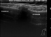
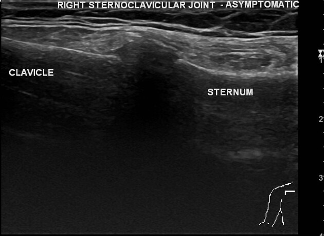
The asymptomatic right sternoclavicular joint was examined for comparison.
Case Discussion
The patient had a left upper chest region contact injury while playing freeze bee. The initial left shoulder radiograph (not uploaded, no copyright) revealed no fracture on the day of injury.
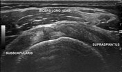
Due to persistent pain, swelling in the left sternoclavicular region, and painful neck and shoulder movements; a shoulder radiograph was repeated which again failed to reveal a fracture. Ultrasound revealed posterior dislocation of the medial end of the left clavicle. The left shoulder ultrasound was normal.
CT angiography was advised to rule out/ rule in upper mediastinal vascular injury. There was no vascular injury.


 Unable to process the form. Check for errors and try again.