Sternoclavicular joint injuries are uncommon and can vary from a mild joint capsule sprain to serious dislocation. This article is focussed on sternoclavicular joint dislocations.
On this page:
Epidemiology
Most cases result from indirect trauma 5, especially high-speed motor vehicle accidents. They can rarely occur in high-impact sports (e.g. American football, rugby) 8. Spontaneous dislocations can also occur, these are typically anterior and occur in young men 2.
Dislocation is rare, accounting for ~2% of joint dislocations and especially when compared to other traumatic upper limb injuries such as clavicular fractures.
Associations
Sternoclavicular joint dislocations are associated with the following injuries 3:
-
anterior dislocation
-
posterior dislocation
subclavian vascular injury
cardiac arrhythmias
tracheal injury
Clinical presentation
Anterior dislocation causes a palpable deformity. Posterior dislocations may be occult on physical examination but, due to compression of thoracic structures, may cause dyspnea, stridor, or dysphagia.
Pathology
The injury can be broadly categorized into two types based on the direction of medial clavicular displacement:
-
anterior
2-3 times more common
less serious
medially-directed impact on the lateral shoulder 8
-
posterior
often occult clinically
can be occult or subtle on radiographs
posteriorly-directed impact on the anteromedial clavicle or anteriorly-directed impacted on the posterolateral shoulder 8
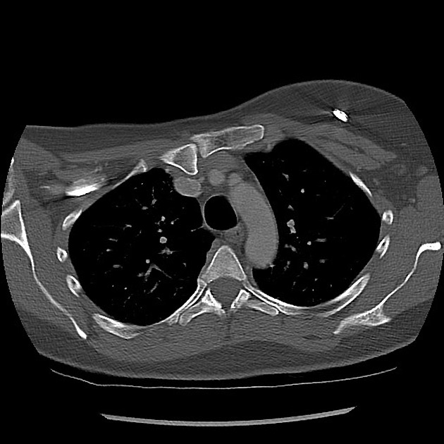
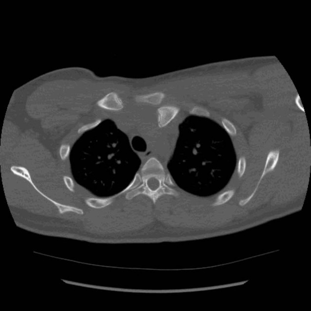
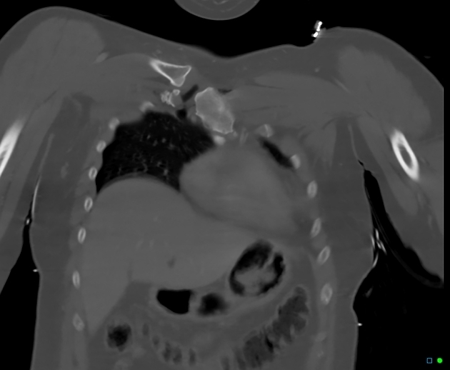
potentially more serious because of the possibility of damage to mediastinal structures (e.g. great vessels, trachea, esophagus, etc.) as a result of posterior displacement of the medial clavicle head
these injuries should prompt assessment of the mediastinal structures with CTA
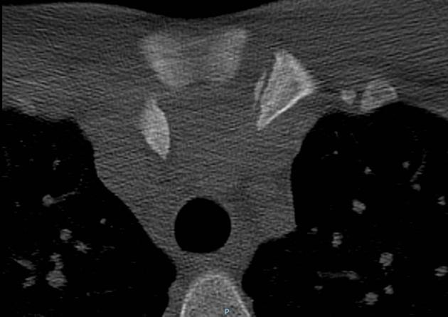
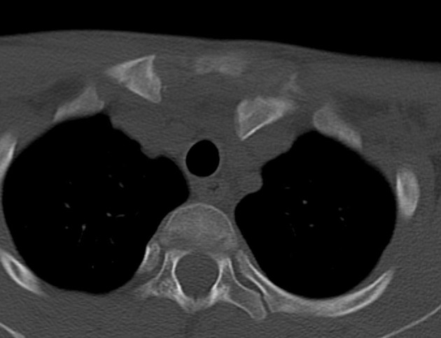
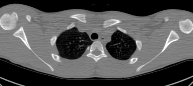
Bilateral dislocations are easily missed on plain film imaging, a rare however necessary consideration, best demonstrated on cross-sectional imaging 7.
Radiographic features
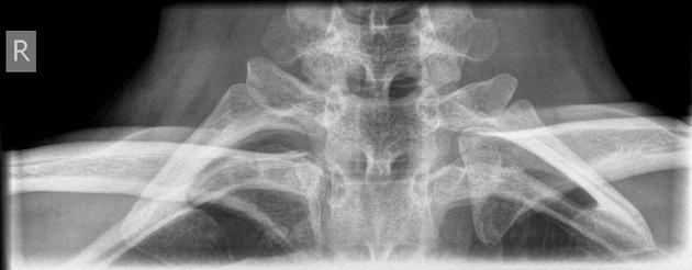
Plain radiograph
joint space widening at the sternoclavicular joint
more easily identified on an angled view, on this view inferior displacement of the medial head of the clavicle is indicative of a posterior dislocation, whereas superior displacement of the clavicle indicates an anterior dislocation 6
difficult to determine anterior or posterior dislocation
Ultrasound
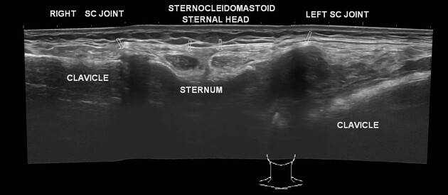
See article: Sternoclavicular joint (ultrasound).
CT
joint space widening and asymmetry at the sternoclavicular joint
associated injuries of the mediastinum
Treatment and prognosis
Options include conservative treatment, especially for anterior dislocation. Posterior dislocations are normally treated with closed reduction. Surgical fixation (ORIF) is usually reserved for unreduced posterior dislocations 2.
Thoracic outlet syndrome may occur as a late complication of posterior dislocation.
Differential diagnosis
posterior sternoclavicular dislocation: medial clavicular physeal fracture in patients <25 years old 8




 Unable to process the form. Check for errors and try again.