
Presentation
Numbness and muscle twitching in the right arm.
Patient Data


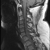
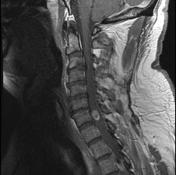


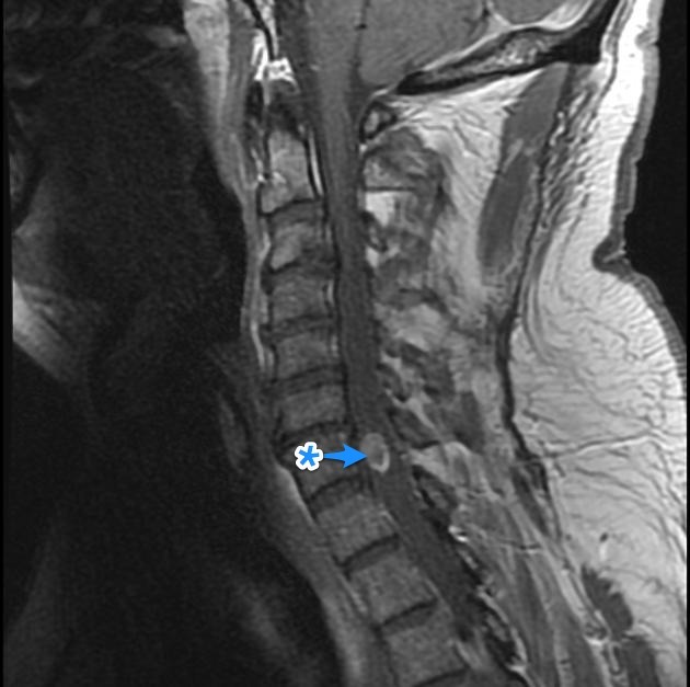
An enhancing intramedullary nodule at C7 is present with extensive perilesional edema. It is an isolated lesion, with no evidence of leptomeningeal disease.
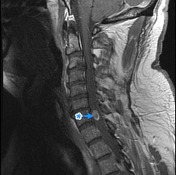
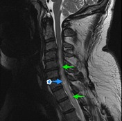
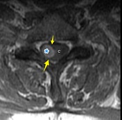
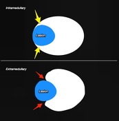
A heterogeneous vividly enhancing nodule ( * ) is associated with extensive cord edema above and below it (green arrows). The nodule appears intramedullary with a visible 'claw sign' - cord (C) is seen wrapping around it (yellow arrows), rather than being pushed away (red arrows) as would be expected in an extramedullary mass.
This patient had had a left upper pulmonary lobe resection for small cell lung cancer 2 years ago, and had extensive metastatic disease elsewhere.
Case Discussion
This case illustrates an fairly typical appearance of intramedullary spinal cord metastatic deposit, a rare occurrence even in patients with widespread metastatic disease.


 Unable to process the form. Check for errors and try again.