
Presentation
Intermittent bilateral anterior cervical soft tissue draining fistulae. Symptoms present since birth.
Patient Data


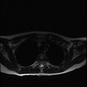
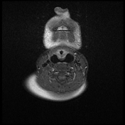
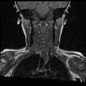
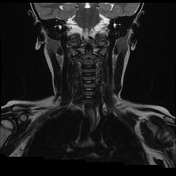
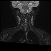
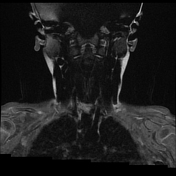
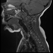
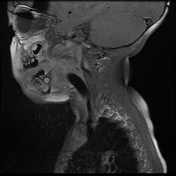
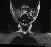
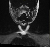
Bilateral fourth branchial cleft fistulae and right branchial cleft cyst. The fistulae originate at the apex of the piriform fossae bilaterally and descend to the base of the neck. The right fistula terminates into tiny palpable right branchial cleft cyst and the left terminates with visible cutaneous opening anterior to the sternocleidomastoid muscle and thyroid gland.
The fistulae extend 45 mm craniocaudally and the right branchial cyst measures approximately 5 mm in diameter.
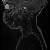
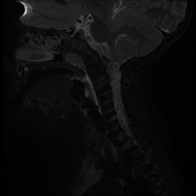
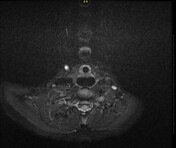
There is no abnormal post-contrast enhancement, excluding superadded sepsis/abscess formation.
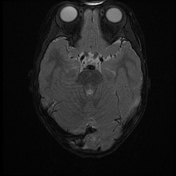
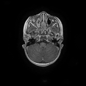
Incidental uncomplicated sinusitis and reactive, benign-appearing, anterior cervical chain lymphadenopathy.
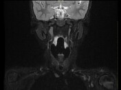
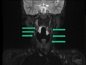
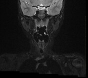
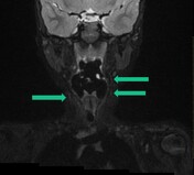
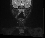
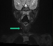
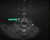
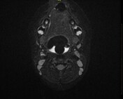
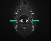
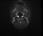
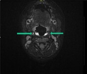
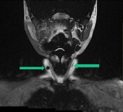
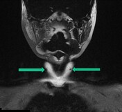
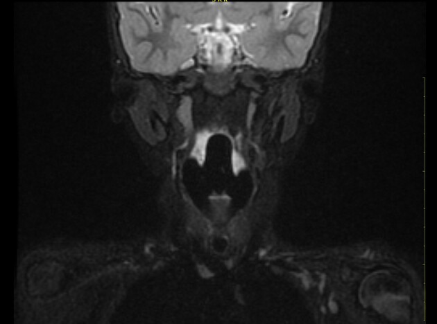
Annotated images demonstrate the craniocaudal course of the bilateral fourth branchial cleft fistulae, extending from the apex of bilateral piriform sinuses and terminating at the right branchial cyst and left cutaneous opening.


Photograph of the anterior cervical soft tissues. The child's neck is in hyperextension.
There are dual cutaneous punctate foci of depigmentation with a left-sided visible fistula opening and palpable tiny right-sided cyst.
The photograph was uploaded with permission.
Case Discussion
Third and fourth branchial cleft anomalies are extremely rare and can be difficult to differentiate radiologically. The relationship of the sinus tract to the superior laryngeal nerve is often determined surgically. The third branchial sinus lies above the superior laryngeal nerve and the fourth branchial sinus lies below it. The third branchial cyst lies posterior to the sternocleidomastoid muscle while the fourth branchial cyst lies anterior to it. The patients often present with recurrent episodes of suppurative thyroiditis/neck abscesses. The fourth branchial sinus may also course deep into the mediastinum and open anteroinferior to the subclavian artery.


 Unable to process the form. Check for errors and try again.