Presentation
Left chest pain after motor vehicle collision.
Patient Data

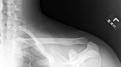
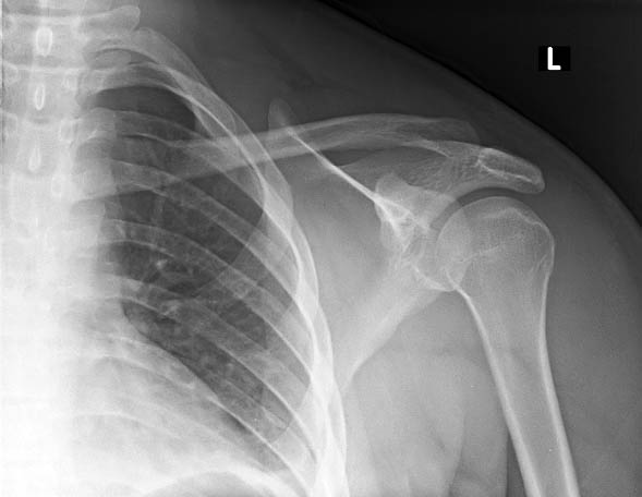
No plain radiographic findings of left clavicle fracture or dislocation.


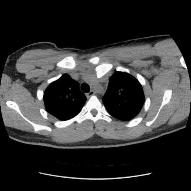
Dislocation left sternoclavicular joint with posterior displacement of the left proximal clavicle. Small anteriorly displaced fracture fragment, but no shaft fracture. Small left pleural effusion with passive atelectasis of the adjacent lung.
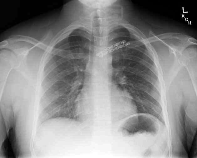
Status post plate and screw fixation left sternoclavicular joint, otherwise normal.
Case Discussion
Plain radiographs may be normal (as in this case) in patient's with a posteriorly dislocated clavicle. CT is much more sensitive. In addition, CT (preferably with intravenous contrast) can assess for mediastinal injuries that may result because of displacement of the clavicle into the mediastinum.


 Unable to process the form. Check for errors and try again.