
Presentation
Abdominal pain and dyspepsia, vomiting, and recent weight loss.
Patient Data
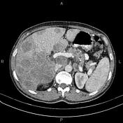


Increased wall thickness due to tumoral infiltration is present at the gastric cardia, subcardia, and proximal of the lesser curvature. The fat plane between the mass and adjacent pancreas is obliterated, and local invasion is suspected.
Multiple enlarged lymph nodes are seen in perigastric, paraceliac, porta hepatis, and para aortic regions with a short axis diameter of less than 20 mm.
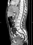
Additionally, numerous ill defined low enhancing masses in the liver less than 100 mm in diameters, accompanied by some perihepatic fluid.
A few nonenhanced simple cortical cysts are seen in both kidneys.
The prostate gland is enlarged.
A small amount of free fluid is present in the pelvis. In addition, in imaged portions of the lower thorax, a small amount of pleural effusion is present on the right side.
Case Discussion
This case demonstrates a gastric mass; pathology proved adenocarcinoma, with suspected invasion into the adjacent pancreas, regional and metastatic enlarged lymph nodes, diffuse hepatic metastasis, and mild ascites.


 Unable to process the form. Check for errors and try again.