
Presentation
Known case of scleroderma with pulmonary manifestations. Follow-up.
Patient Data
Age: 18 years
Gender: Female
From the case:
Scleroderma - pulmonary manifestations


Download
Info
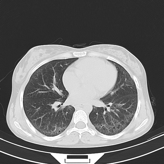
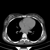
Peripheral ground glass and reticular opacities, microcystic honeycombing, and scattered small air cysts, with relative immediate subpleural sparing.
The presence of said findings in the anterolateral upper lobes and posterosuperior lower lobes (four corners sign 1) is a specific for scleroderma-associated interstitial lung disease.
Scattered tiny lung nodules.
Dilated esophagus.
Case Discussion
This is a case of interstitial lung disease associated with systemic sclerosis, with a pattern highly suggestive of non-specific interstitial pneumonia (NSIP).


 Unable to process the form. Check for errors and try again.