Presentation
Right thigh swelling for 6 months, increasing in size. Associated with constitutional symptoms (weight loss) and night sweat.
Patient Data
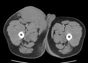


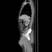

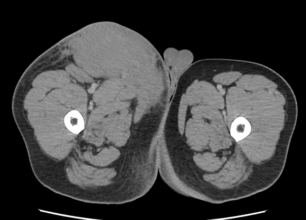
Multiple homogenously enhancing soft tissue masses at the right external iliac region, right inguinal region, and right anterior and medial thigh compartments. These soft tissue masses are in close proximity to the right lower limb vessels. The masses are located in the subcutaneous tissue layer. The external iliac and femoral vessels are well opacified. The masses abut the medial compartment muscles of the right thigh.
Short segment of small bowel-small bowel intussusception at the left iliac fossa where the small bowel walls appear thickened. No other obvious bowel-related mass. No bowel loops dilatation.
No abdominal lymphadenopathy.
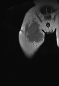

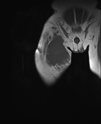

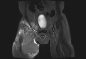

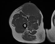


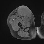


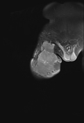

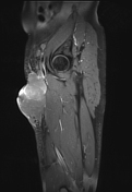

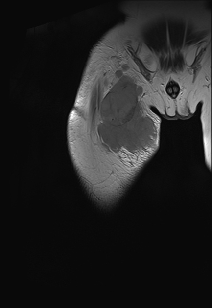
Multiple subcutaneous masses at the right inguinal region extend into both right thigh anterior and medial compartments. They demonstrate hyperintense on T2/STIR sequences and isointense on T1. On post gadolinium administration, these masses show minimal enhancement.
Posteromedially, the mass abuts the right pectineus muscle, right adductor longus and right gracilis muscles with muscle sheath enhancement.
A clear fat plane between the masses and the rest of the adjacent thigh muscles. No clear evidence of muscle infiltration.
The femoral neurovascular bundle is not involved. The adjacent femoral vessels preserved their normal flow void signal.
Multiple enlarged external iliac, right inguinal and right anterior thigh lymph nodes.
After 3 cycles of chemotherapy, the patient presented with sudden onset of abdominal pain. The clinical examination revealed peritonism. Urgent contrast-enhanced CT abdomen was performed to rule out the possibility of bowel perforation, especially the previously noted small bowel intussusception.
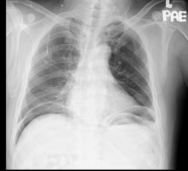
Large amount of free air under both diaphragms in keeping with pneumoperitoneum.
Right port.
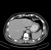

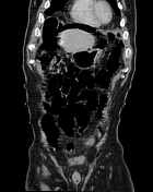

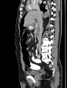

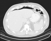

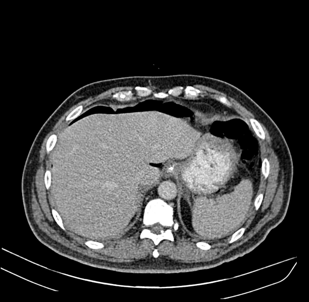
Large pneumoperitoneum, predominantly at the upper abdomen and bilateral subphrenic regions.
Accumulation of free fluid at the left subphrenic region. No obvious oral contrast extravasation into the peritoneum.
Minimal free fluid at the right paracolic gutter and pelvis.
No obvious bowel-related mass or bowel loops dilatation. The previously noted small bowel intussusception at the left iliac fossa has resolved.
The appendix is air-filled, normal in caliber and retrocecal in position. No periappendiceal fat streakiness.
The previously noted right inguinal and external iliac lymphadenopathy have largely resolved. Residual lymph node at the right inguinal region.
Mild degree of left pleural effusion with adjacent passive lower lobe atelectasis.
Minimal right lower lobe consolidation.
Case Discussion
Right inguinal and external iliac masses are suspicious of lymphadenopathy. Proceeded with trucut biopsy of the masses.
Trucut biopsy right medial thigh mass histopathology result:
Microscopic: The section shows multiple fragments of fibromuscular tissue diffusely infiltrated by malignant lymphoid cells. The malignant cells are medium in size, characterized by hyperchromatic to vesicular nuclei with fine chromatin, some with conspicuous nucleoli, and scanty cytoplasm. Mitosis and apoptotic bodies are easily seen. immunohistochemical studies show the malignant lymphoid cells are positive for CD20, CD10, and BCL6, and negative for CD3, MUN-1, BCL2, and TdT. The proliferation index, Ki67 is 100%.
Trucut biopsy of right medial thigh mass: Consistent with Burkitt's lymphoma.
Small bowel intussusception in the adult population usually has a lead point, with more frequent identifications in up to 90% of adult cases. In this case, it is due to Burkitt's lymphoma.
Post chemotherapy, the primary lymphadenopathy has largely resolved in keeping with treatment responsive. The previously noted small bowel intussusception has resolved as well.
The patient had an emergency laparotomy where a perforated gastric ulcer (0.5cm) at the proximal body of the stomach was found intraoperatively and thus received a Modified Graham patch. The pre-operative CT scan showed that pneumoperitoneum is largely confined to both subphrenic regions with loculated free fluid in the left subphrenic region. It has raised suspicion that the site of perforation was at the fundus or body of the stomach, though no active oral contrast extravasation in the CT scan was seen, likely due to the non-gravity-dependent location of the perforated gastric ulcer.


 Unable to process the form. Check for errors and try again.