
Presentation
Painless mobile right submandibular region soft swelling for one year.
Patient Data

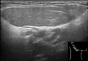
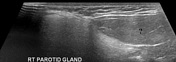
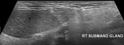
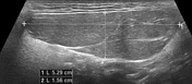
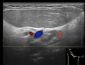
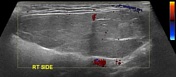
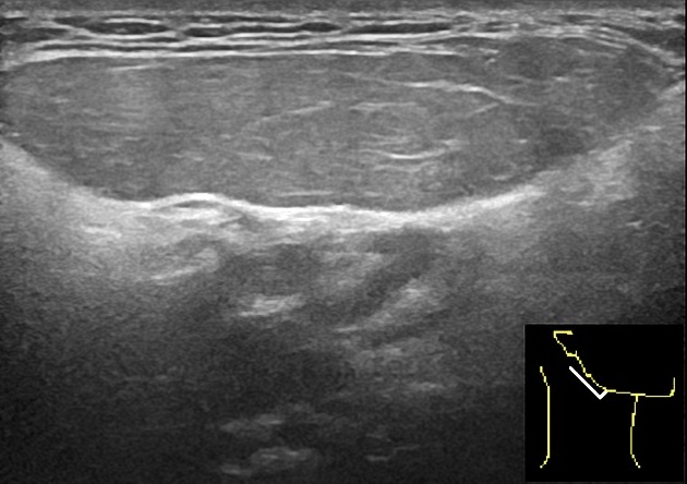
Well defined homogeneous oval shaped capsulated fat echogenicity lesion measuring about 1.5 x 5.3 cm in the right submandibular region between the parotid and submandibular glands. No vascularity is seen in it on color Doppler ultrasound examination.


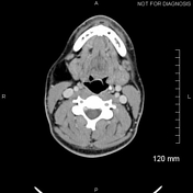
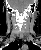
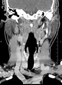
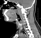
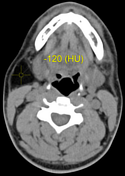
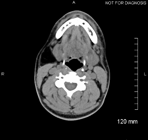
Fat density lesion (average density of -120 HU) in the right upper neck just below the parotid gland, along the posterolateral aspect of the submandibular gland. No calcifications, solid component or enhancement is seen in it. No compression effects are seen on the adjacent structures. Gross morphology of the parotid and submandibular salivary glands is within normal limits. Multiple small sub centimeter lymph nodes, likely reactive in nature, are seen on both sides of the neck.
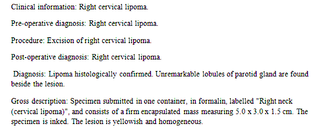
The lesion was completely excised and the histopathology showed lipoma.
Case Discussion
Imaging features as well as the histopathology are consistent with lipoma in the right upper neck.


 Unable to process the form. Check for errors and try again.