
Presentation
High school athlete with acute onset of left hip pain during track and field competition.
Patient Data

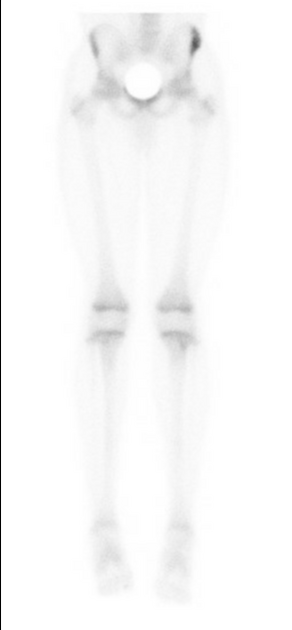
Single-phase bone scan of the lower body obtained 10 days after onset of symptoms demonstrates abnormal increased radiotracer at the left anterior superior iliac spine (ASIS). This is asymmetric from the right ASIS. Bony uptake is otherwise physiologic.

Frontal x-ray of the pelvis obtained day after onset of symptoms shows no acute abnormality.
Case Discussion
Bone scan imaging agents are phosphate analogs. They localize to bone in proportion to osteoblastic activity at sites of bony remodeling and, to a lesser degree, also in areas of increased blood flow due to elevated delivery of the radiotracer.
The clinical history of acute onset of left hip pain during a track and field competition is suggestive of a hip or pelvic avulsion injury. The normal pelvis x-ray excludes a displaced fracture or unsuspected bony lesion. The abnormal radiotracer uptake on subsequent bone scan, localizing to the anterior superior iliac spine (ASIS), is most consistent with a non-displaced ASIS avulsion injury.


 Unable to process the form. Check for errors and try again.